Splanchnology Cremona Key Info – Alimentary System, University Notes
Document from University about Splanchnology Cremona Key Info – Alimentary System. The Pdf provides key information on the alimentary system, covering the anatomy of the mouth, stomach, and their vascular and lymphatic systems, useful for Biology students.
See more45 Pages

Unlock the full PDF for free
Sign up to get full access to the document and start transforming it with AI.
Preview
Alimentary System Overview
- The alimentary system is the main system through which the absorption of nutrients required for energy intake from food takes place.
- It can be seen as a large duct (indeed called alimentary canal) in which a series of organs involved in secretion, absorption, and excretion are found.
- Each organ of the alimentary system is identified by a particular lining epithelium and intramural glands which help define its function within the system.
- Extramural glands are examples of glands/organs that secrete exocrine factors into other organs to permit their full function. Examples of these are the primary glands of the mouth, identified as the parotid, sublingual, and submandibular glands, and the liver and pancreas for their factor secretion in the duodenum.
. It is important to note the topographic relationships of the alimentary canal, where in the head are located the mouth, the isthmus of the fauces, and a part of the pharynx, in the neck the remaining part of the pharynx and the first portion of the oesophagus, in the thorax the middle part of the oesophagus, in the abdomen the final portion of the oesophagus, the stomach, liver, pancreas, gallbladder, duodenum, jejunum, ileum, cecum, ascending colon, transverse colon, and the descending colon, and in the pelvis the final part of the large intestine, highlighted by the sigmoid colon, and the first part of the rectum.
. It is important to note that all organs within the abdominal cavity (except the pancreas and part of the duodenum) are covered by a layer of serosa called peritoneum. The pancreas and part of duodenum are instead referred to as retroperitoneal, whereas organs wrapped in peritoneum are peritoneal organs.
Mouth Anatomy
- The mouth is the first portion of the alimentary canal. It possesses teeth which are the primary organs for mastication as well as the tongue, which is responsible for speech and taste perception.
- The mucosa of the mouth cavity is continuous throughout the entire structure, except at the level of the neck of the teeth. The mucosa is characterised by a series of innervations to allow for differential perception (highlighted by the innervation of 5 total cranial nerves).
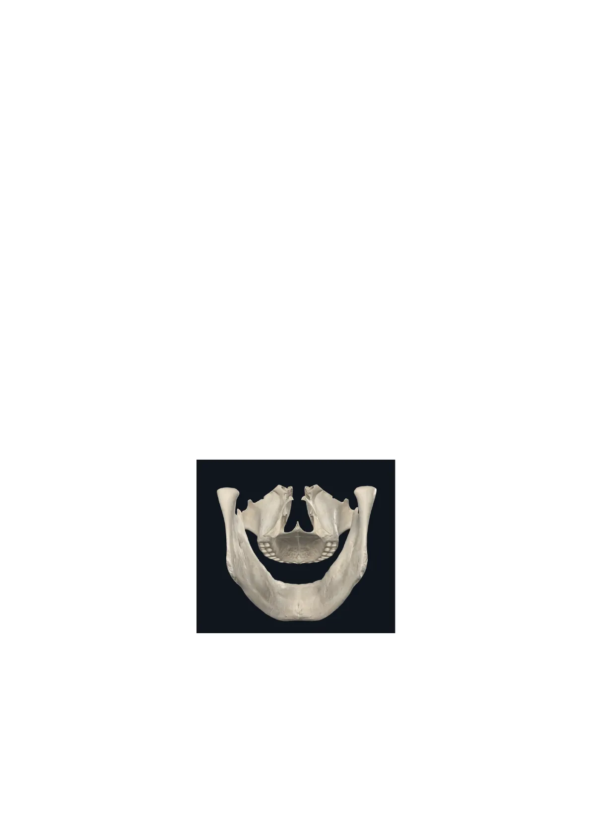
- The mouth cavity possesses a skeletal scaffold primarily highlighted by the left and right maxillary bones superiorly and anteriorly, superiorly in the mouth cavity, posterior to the maxillary bones, are the palatine bones, and inferiorly the mandible, which forms the skeletal scaffold of the chin and inferior layer of teeth.
. On top of the skeletal scaffold are expandable layers composed by the lips, cheeks, floor of mouth, and soft palate.
- The mouth cavity is divided into 2 distinct sections by the alveolar arches and dental arcades, specifically anteriorly and externally of the alveolar arches and dental arcades is the oral vestibule, and posterior to these is the oral cavity proper. The oral vestibule and oral cavity proper are connected to one another via the retromolar triangle.
Oral Vestibule
· The oral vestibule constitutes anteriorly by the lips, posteriorly by the dentoalveolar arches, and laterally by the cheeks. When the mouth is relaxed, the oral vestibule has a sort of horseshoe shape. It is bound superiorly and inferiorly by the vestibular sulci, which are grooves where the mucosa of the lips transitions to the gingival and alveolar mucosa.
- The gingiva wraps around the neck of the tooth and on the vestibular side proceeds to occupy also the interdental spaces.
- The ducts of labial and buccal glands (minor salivary glands) open into the vestibule and the parotid duct. The vestibule communicates with the oral cavity proper by means of the retromolar triangle and the interdental spaces.
Oral Cavity Proper
. The oral cavity proper is the second compartment of the mouth cavity and is delimited anteriorly and laterally by the dentoalveolar arches, superiorly by the hard palate, posteriorly by the isthmus of the fauces and the soft palate, and inferiorly by the floor of the mouth.
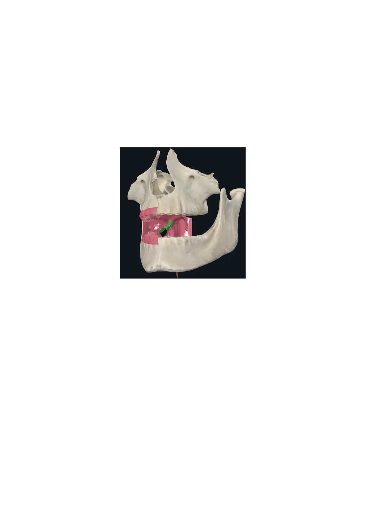
- The oral cavity proper communicates extensively with the oral vestibule, especially when the mandible is lowered. It is important to note that the isthmus of the fauces is a short and narrow space that connects the oral cavity with the oropharynx, and is delimited by the palatoglossal folds (seen in green).
- The mouth cavity possesses a major large opening with which it communicates with the outside, called oral orifice.
Lips Structure
- The lips are musculomembranous structures located between the nose, chin, and cheeks, that limit the oral fissure.
- Lips have an internal and external surface, as well as a free border. The internal surface is in contact with the gingivodental arches, where indeed they make a forward groove. The external surface is instead delimited in two different ways for the upper and lower lip.
- The external surface of the upper lip is delimited superiorly by the base of the nose, laterally by two sulci called nasolabial sulci, which develop from the alae of nose oblique downwards to separate the upper lip from the cheeks, and medially below the nose there is the philtrum, which is another sulcus which goes from the nasal septum to the free border of the upper lip, delimiting a small portion referred to as tubercle of the upper lip.
- The external surface of the lower lip is instead delimited inferiorly by a concave (oriented downwards) sulcus called mentolabial sulcus. The free borders of both lips represent the site where the skin of the external surface flips into the internal surface, transitioning into labial epithelium. We also note a change in colour, where the internal surface is much shinier, smoother, and more pinkish in colour, due to the increased thinness of the epithelium and the underlying large vascularisation of the papillary layer of the dermis.
Lip Vascularization
- The upper and lower lips are vascularised by the superior and inferior labial arteries, respectively, which are branches of the facial artery, and are drained by the superior and inferior labial veins, which are tributaries of the facial vein and submental vein, respectively.
Mandible and Temporomandibular Joint
Lateral view Joint capsule Lateral (temporomandibular) lig. Styloid process Stylomandibular lig. Sphenomandibular lig. Sphenomandibular lig .- (phantom) Jaws closed Mandibular fossa Articular disc Articular tubercle Jaws widely opened (hinge and gliding actions combined) Jaws slightly open (hinge action predominates) & Notes
- The mandible is composed of a body and 16 alveolar sockets, which house the teeth. Posterior to the body of the mandible is the ramus, a flattened portion of membranous bone. The ramus terminates in two distinct processes:
- The anterior process, known as the coronoid process, serves as the insertion point for the temporalis muscle.
- The posterior process, also called the condylar process, comprises the head (articulating with the temporal bone at the temporomandibular joint) and the neck, which provides attachment for the lateral pterygoid muscle.
- The temporomandibular joint (TMJ) includes the mandibular fossa, which is part of the temporal bone and lined by fibrocartilage (not hyaline cartilage, as fibrocartilage is specific to this joint). The TMJ allows for a wide range of movements essential for activities such as talking and chewing.
- A sudden movement or displacement of the articular disc within the TMJ can produce a clicking sound, a common occurrence in certain jaw disorders. The articular disc, composed of fibrous cartilage, is sometimes referred to as the meniscus.
- The TMJ is stabilised by three main ligaments:
- The sphenomandibular ligament (internal), connecting the sphenoid bone to the mandible.
- The lateral temporomandibular ligament (external), providing lateral reinforcement.
- The stylomandibular ligament (posterior), extending between the styloid process of the temporal bone and the mandible.
. Additionally, the styloid process serves as the origin point for three muscles: the styloglossus, stylohyoid, and stylopharyngeus.
Palate Structure
External ptery- goid plate Internal ptery- goid plate Horizontal portion of palate bone Pyramidal proc. of palate bone Transverse pala- tine suture Posterior nasal spine Lesser palatine foramina Greater palatine foramen Zygomatic process of maxilla Palatine grooves Palatine spines Incisive suturé Palatine process of maxilla Incisive foramen Median palatine suture
- The palate is separated into hard and soft palate, where the anterior two thirds correspond to the hard palate and the posterior third to the soft palate.
Hard Palate
- The hard palate forms the superior wall of the oral cavity proper, and is delimited anteriorly and laterally by the alveolar processes of the maxillary bones. Posteriorly instead is delimited by the soft palate and the isthmus of the fauces.
- The two maxillary bones attach together via the median palatine suture, between the two palatine processes of the maxillary bones. Here in the midline is found the palatine raphe, which splits the hard palate into two symmetrical halves. In the anterior portion of the palatine raphe is the incisive foramen, where veins draining the hard palate pass through to drain into superior nasal branches.
- The skeletal scaffold of the hard palate is therefore constituted superiorly anteriorly by the median palatine suture between the palatine processes of the two maxillary bones, and superiorly posteriorly by the transverse palatine suture, made between the two palatine bones.
- The arterial supply of the hard palate is mainly mediated by the sphenopalatine artery and the descending palatine artery. These are both branches of the maxillary artery. The descending palatine artery is the major vascular supply, and once in the hard palate splits into lesser palatine arteries and greater palatine arteries, based on the caliber of the vessels themselves.
- The venous drainage is satellite to the arterial vascularisation, where veins either go up through the pterygomaxillary fixture to join the pterygoid plexus, or go up the incisive foramen and rejoin veins draining the nasal mucosa.
Soft Palate
- The soft palate is the posterior continuation of the hard palate, and represents the posterior third of the palate structure. Its anterior margin is indeed the posterior surface of the posterior third of the hard palate, whilst its posterior surface is free. The lateral margins proceed with the gingivae and later with the walls of the pharynx.
Can’t find what you’re looking for?
Explore more topics in the Algor library or create your own materials with AI.
