Excretory System: Anatomy and Function of Kidneys and Nephrons
Slides about Excretory System. The Pdf provides a detailed overview of the excretory system, with a focus on the kidneys and nephrons. This Biology material for High school students explains how nephrons filter blood to produce urine, describing the different parts of the kidney and the components of the nephron.
See more11 Pages

Unlock the full PDF for free
Sign up to get full access to the document and start transforming it with AI.
Preview
EXCRETORY SYSTEM NUTRITION
OBTAINING
Digestive system
Nutrients
Respiratory
system
Oxygen
DISTRIBUTING
Circulatory
system
Nutrients and oxygen
and collecting waste
products and carbon
dioxide
ELIMINATING
Excretory system
Waste products
Respiratory
system
Carbon dioxide
Excretory Products and Organs
What are the excretory products
of the various excretory organs?
EXCRETORY SYSTEM
Excretion is the elimination of waste products which result from the metabolic activity of cells.
Without excretion, the waste products would accumulate and become toxic.
Liver
Urinary system
Respiratory system
Sweat glands
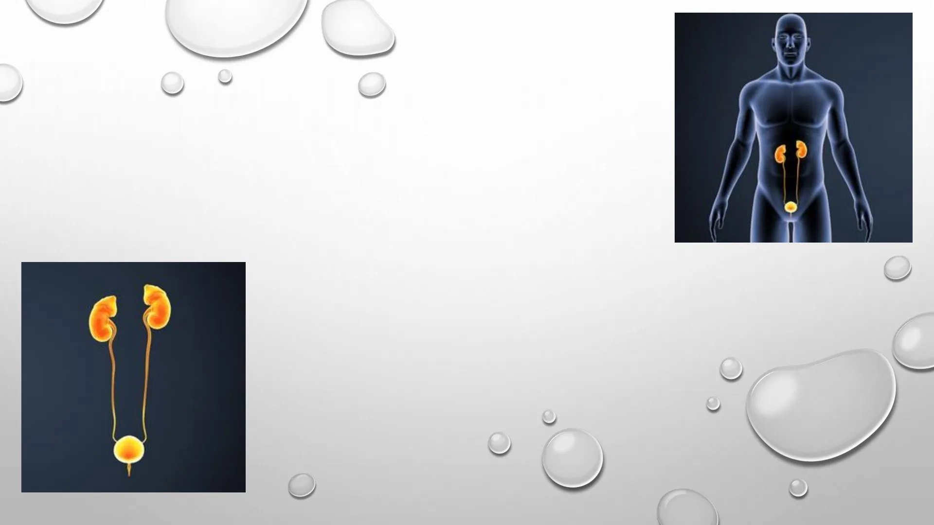
THE URINARY SYSTEM
Adrenal gland
Kidneys
Adrenal glands - Endocrine system
These endocrine glands secrete
hormones such as adrenaline and
corticosteroids.
Renal
vein
Renal
artery
Inferior vena cava
Aorta
Urinary bladder
Ureters
Urethra
The renal artery enters and
the renal vein leaves the
kidneys on its concave side.
Include the draw in
your notebook.
Include the draw in
your notebook.
KIDNEYS
Cortex: outer layer
Medulla
Renal pelvis
The renal pelvis is the
innermost part of the
kidney. It is funnel-shaped
and connected to the ureter.
The medulla is the striated middle layer of the
kidney. This is split into triangular sections
called renal pyramids or Malpighi's pyramids.
Kidney
Unfiltered
blood
Filtered
blood
Ureter
Urine exits
to bladder
Each kidney contains approximately one million
nephrons. These units filter the blood and produce urine.
Nephron Structure
Nephron
Glomerulus
Filtered
blood
Tubule
Unfiltered
blood
Collecting duct
Urine exits duct
Glomerulus
Filtered
blood
Bowman's capsule
Distal convoluted
tubule
Unfiltered
blood
Proximal
convoluted tubule
Cortex-medulla
junction
I
-
Venule
Collecting duct
Loop of
Henle
Peritubular
capillary
network
Include the draw in
your notebook.
Urine flows into renal pelvis
NEPHRONS
Bowman's capsule is a hollow, cup-shaped sac
surrounding a ball of capillaries called the
glomerulus. This structure is inside the renal
cortex.
Convoluted tubules are twisted tubes. They may
be proximal convoluted tubules running from
Bowman's capsule to the loop of Henle or distal
convoluted tubules running from the loop of
Henle to the collecting duct.
The loop of Henle is a U-shaped tube located
between the proximal and distal convoluted
tubules. This part of the nephron enters the
medulla of the kidneys.
URINARY TRACT
Ureters
They are two 25 cm-long, narrow tubes.
They take urine from the kidneys to the urinary bladder.
Ureter
Smooth muscle
layers
-Internal sphincter
External sphincter
Urinary bladder
Expandable, muscular organ. Up to
half a litre of urine collects here,
although it can store up to two litres.
Urethra
It connects the urinary bladder with the
outside and has two sphincters to control
the output of urine. The internal sphincter is
under involuntary control while the external
one is under voluntary control.
right kidney
left kidney
ureters
bladder
urethra
1
Bowman's capsule
Filtered
blood
Distal convoluted
tubule
Unfiltered
blood
2
K+
3
Proximal
convoluted tubule
Cortex-medulla
junction
I
-
Venule
Collecting duct
2
Loop of
Henle
Peritubular
capillary
network
Urine flows into renal pelvis
4
THE EXCRETORY PROCESS
- Absorption: liquid passes from the blood in
the glomerulus into the Bowman's capsule. This
process filters out water, waste products (urea),
vitamins, amino acids, mineral salts and glucose. - Reabsorption: substances such as water,
glucose, amino acids and mineral salts are
returned to the blood from the tubules and the
loop of Henle. - Some substances, such as potassium ions (K+),
pass from the interstitial fluids into the final
part of the nephron. - Urine is produced and flows to the renal pelvis.
THE EXCRETORY PROCESS
- Urine flows continuously from the ureters into the
bladder. - Urination: when the bladder is full, urine flows
through urethra from the bladder to the outside of the
body. This process is controlled by two small muscles
called sphincters at the top and bottom of the urethra.
Kidney
Kidney
(inside
view)
Renal
pelvis
Ureter
5
Bladder
Opening of
ureter
(ureteral orifice)
Internal
sphincter
Urethra
External
sphincter
6
THE URINE
Composition of Urine
Urine is a clear, yellow liquid composed of:
- 95% water.
- 3% of toxic substances, such as urea and uric
acid. - 2% of mineral salts, mainly sodium and
potassium.
Functions of Urine
- Eliminate toxic waste from the body.
- Regulate the levels of ions in the blood.
- Balance fluid levels.
- Regulate blood volume and pressure.
What does urea
come from?
Can’t find what you’re looking for?
Explore more topics in the Algor library or create your own materials with AI.
