Lecture #1: Neurohistology, Midwestern University Presentation
Slides from Midwestern University about Lecture #1: Neurohistology. The Pdf explores the organization of the nervous system, distinguishing between central and peripheral systems. This University Biology material details neuron parts, axonal transport, and synaptic components.
Ver más12 páginas

Visualiza gratis el PDF completo
Regístrate para acceder al documento completo y transformarlo con la IA.
Vista previa
Neurohistology Learning Objectives
- Be able to differentiate the blood-CSF barrier from the blood-brain barrier.
- Be able to list the major cell types of the nervous system and their characteristics.
- Be able to describe the overall structure of the typical neuron and the appearance and function of the major components of a neuron.
- Be able to describe the overall structure of a synapse, as well as the specific structure and function of the major components of a synapse.
- Be able to list the special characteristics of neurons that subserve the function of communication.
- Be able to list the major functions of each type of glial cell.
Organization of the Nervous System
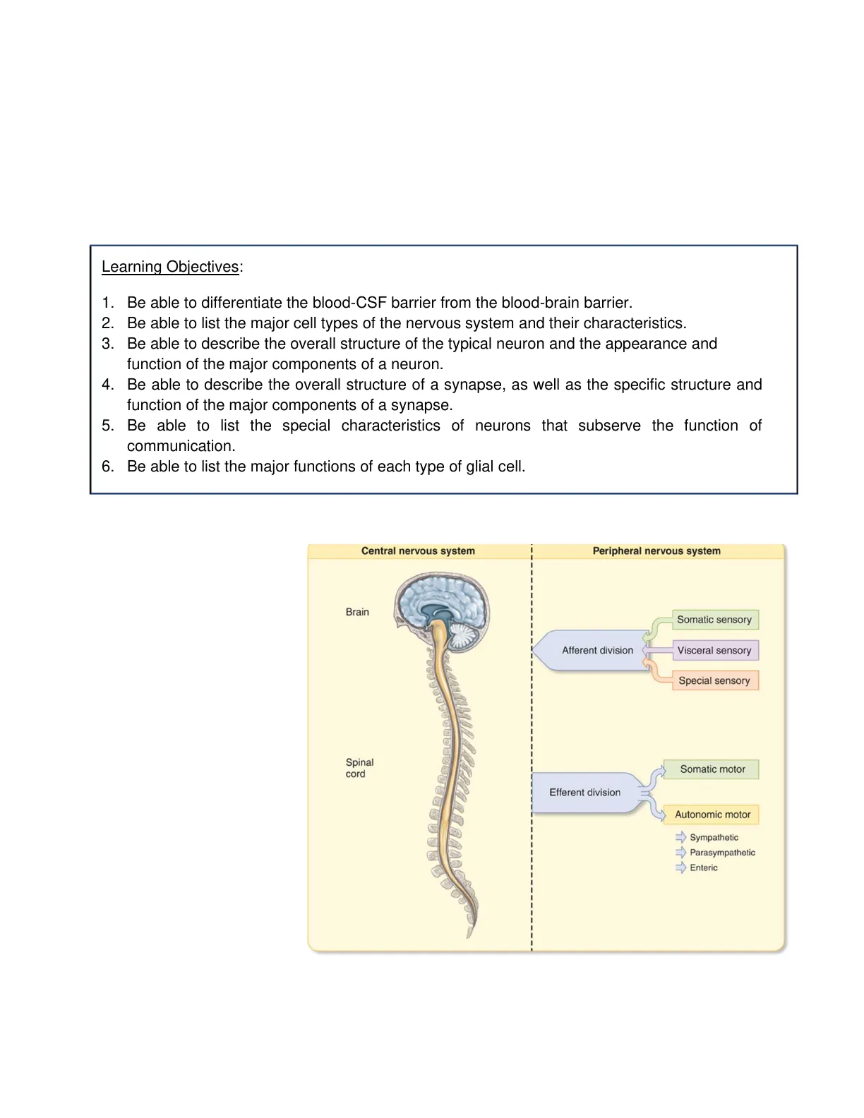
The nervous system is divided into the central nervous system (CNS) and the peripheral nervous system (PNS). CNS includes the brain and spinal cord. The PNS consists of sensory (afferent) neurons that transmit sensory information to the CNS, and efferent neurons that carry signals from the central nervous system to the target cells (Fig.1). The CNS is encased within the bone (skull). The spinal cord goes through a canal through the vertebrae.
Central nervous system Peripheral nervous system Brain Somatic sensory Afferent division Visceral sensory Special sensory Spinal cord Somatic motor Efferent division Autonomic motor Sympathetic Parasympathetic Enteric Fig. 1. Central and Peripheral Nervous System. Lecture #1 - Neurohistology p. 1-1
Choroid Plexus and Ventricular System
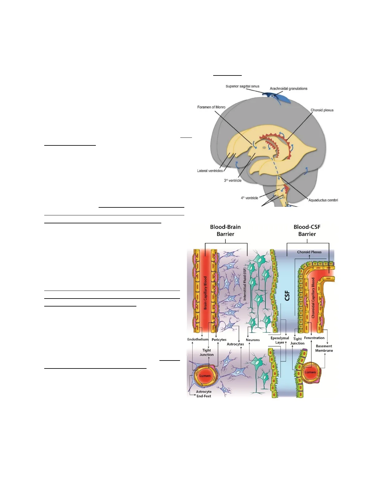
The entire nervous system is bathed in a colorless fluid called cerebrospinal fluid (CSF) created by the choroid plexus, which is the tissue that lines the ventricles (hollow spaces) in the brain. The choroid plexus is a complex network of capillaries lined by specialized cells. CSF offers a unique chemical environment as well as an Superior sagittal sinus Arachnoidal granulations additional physical protection layer. It flows in and around the brain and spinal cord to help cushion them from injury and provide nutrients.
Blood-CSF Barrier (BCSFB)
Foramen of Monro Blood-CSF barrier (BCSFB): Inside the ventricular system of the brain, we find four choroid plexuses, one in each of the two lateral ventricles, one in the third, and one in the fourth ventricle. They have two major functions: to produce CSF, as mentioned above, and to serve as a barrier in the brain separating the blood from the CSF, known as the blood-CSF barrier (BCSFB). This barrier is composed of cells with tight junctions that limit the passage of substances between the cells. The mechanism of the blood- CSF barrier is a well-studied target for the delivery of novel pharmacologic agents.
Blood-Brain Barrier (BBB)
BBB is formed by the endothelial cells of the brain microvasculature, which are connected by tight junctions. Like the blood- CSF barrier (BCSFB), the BBB barrier promotes the exchange and elimination of metabolites while blocking the entry of blood- borne chemicals into the brain. However, the substances exchanged between each respective barrier differ significantly due to their distinct functions. Drugs can cross the barrier simply by passive diffusion if they are sufficiently lipid soluble (or have been made lipophilic by appropriate chemical modifications). Carrier protein-mediated active transport can allow many essential compounds, such as glucose and amino acids, to enter the endothelial cytoplasm and then be released into the brain. The tight junctions present in the endothelium and all the elements of the neurovascular unit contribute to the high selectivity of the BBB.
Choroid plexus Lateral ventricles 3ª ventricle 4th ventricle Aquaductus cerebri Fig. 2. Organization of the ventricular system of the brain Blood-Brain Barrier Blood-CSF Barrier Choroid Plexus Brain Capillary Blood Interstitial Fluid (ISF) CSF Choroidal Capillary Blood she Endothelium Pericytes Neurons 4 Ependymal Tight Fenestration Layer + Junction Astrocytes Basement Membrane Tight Junction Lumen Lumen Astrocyte * End-Feet Fig. 3. Blood-CSF Barrier and Blood-Brain Barrier Lecture #1 - Neurohistology p. 1-2
Although in principle both the BCSFB and BBB serve the same defensive purpose for the CNS, their distinct structure allows the interchange of different substances between the bloodstream and brain. Unlike the BBB, the capillaries of the blood-CSF barrier are relatively leaky and permeable to small molecules, thus allowing, among other processes, the rapid delivery of water through the bloodstream to the surrounding epithelial cells for the CSF production in the choroid plexus.
Cell Types in the Nervous System
Histologically, the nervous system can be divided into two broad categories: nerve cells or neurons, and non-neuronal cells.
Neuronal Cells
. Neurons: The nerve cell, or neuron, is the basic building block of the nervous system. Neurons have extended processes that project from the nerve cell body. Dendrites, which receive incoming signals, and axons, which transmit information, are examples of these processes. Axons and dendrites are crucial components of how neurons communicate with one another and with other cells.
Non-Neuronal Cells
In the central nervous system, non-neuronal cells outnumber neurons. These many cells are essential for neuronal survival and maintenance, but they also actively contribute to brain growth and function.
- Glial cells: Usually referred to as glia or neuroglia, provide support for neurons. The word glia is Greek for "glue" and reflects the previous belief that these cells held the nervous system together. Glial cells are not directly involved in synaptic transmission or electrical signaling, but they do communicate with neurons and provide structural and biochemical support.
- Other cell types:
- Meningeal cells: The meninges are the fibrous layers that cover and protect the CNS. Within its three layers (dura, arachnoid, and pia), the meninges include a remarkably diverse range of cell types, such as fibroblasts, a complex lymphatic system, and immune cells different from the central nervous system.
- Epithelial and endothelial cells: These are constituents of the choroid plexus and the blood-brain barrier.
In the most general sense, there are two general types of tissue in the CNS:
- Gray matter and white matter.
Lecture #1 - Neurohistology p. 1-3
- Gray matter is composed of nerve cell bodies and dendrites.
- White matter is mostly composed of axons, which are covered in myelin, giving it its characteristic white appearance.
Gray matter White matter Myelinated axon Oligodendrocyte Dendrite Cell body Axon terminal of presynaptic cell Fig. 4. Gray matter and white matter
Parts of a Neuron
Neurons are the structural and functional unit of the nervous system. The nervous system contains several different kinds of neurons, yet they all have some characteristics in common. Typically, the neuron has a cell body, an axon, and dendritic processes that arise from the soma.
Dendrites (receptive regions) Cell body (biosynthetic center and receptive region) Neuron cell body Nucleus (a) Dendritic spine Nucleolus Axon (impulse generating and conducting region) Impulse direction Node of Ranvier L Axon hillock Schwann cell (one inter- node) Axonal terminais (secretory component) Neurilemma (sheath of Schwann) Terminal branches (telodendria) Fig. 5. Neuron morphology
Cell Body (Soma)
The cell body is the control center. It has a nucleus for protein transcription and translation, and organelles needed to direct cellular activity, except for centrioles, which are not needed in amitotic cells. In comparison to the rest of the cell, the soma is typically small and contains a nucleus, Golgi apparatus, mitochondria, and Nissl bodies that consist of rough endoplasmic reticulum and ribosomes. All cytoskeletal proteins are produced on free polysomes (a group of two or more consecutive ribosomes) in the cell body and then transported to their different cellular compartments. Lecture #1 - Neurohistology p. 1-4 Nissi bodies
The neuron has an elaborate cytoskeleton that consists of tubulin that forms microtubules, neurofilaments, and microfilaments. The cytoskeleton orchestrates many functions, such as axonal and dendritic growth, the trafficking of membrane components, organelles, and vesicles, and their appropriate location.
Dendrites
Dendrites are processes that extend outward from the cell body. The function of the dendrites is to receive incoming electrical signals sent by other neurons. They contain many ribosomes and specific cytoskeletal proteins. The dendritic arborization varies in size and branching and is crucial in establishing the number of inputs that a specific neuron receives. Neurons with dense dendritic branches are innervated by many neurons, which promotes a large integration of information. The dendrites can increase the surface of communication with the presence of dendritic spines, which are small dendrite membrane protrusions that are of critical importance in learning and memory.
axon terminal (from different neuron) dendrites nucleus spines cell body (soma) 1um axon Fig. 6. Spines in dendrites. Axons provide synaptic signals to dendrites, and the sum of these signals determines whether the neuron will fire an action potential.
Axon
The axon carries outgoing information to the target cell. Most neurons have one axon. The axon lacks free ribosomes and rough endoplasmic reticulum after the initial segments, and because it contains protein channels, the axonic membrane (axolemma) differs from the somatic membrane in terms of composition. Axons can have sheaths made of an insulating substance called myelin. The length of the axon varies from a few microns to over a meter.
- Important axonal parts: Axon hillock: The action potential generated in the axon hillock propagates along the axon and causes the release of the neurotransmitter into the synapse, which allows the neuron to communicate with another cell. Axon terminal: The distal part of the axon that contains synaptic vesicles. Note that efferent neurons have axon terminals, and many autonomic neurons also have enlarged regions along the axon called varicosities. Growth cone: This is a specialized structure at the tip of the axon, very mobile, that directs the axon's growth. It has many microtubules and actin filaments. Collaterals: The axonal branches are known as axon collaterals.
Lecture #1 - Neurohistology p. 1-5
¿Non has encontrado lo que buscabas?
Explora otros temas en la Algor library o crea directamente tus materiales con la IA.
