Unit 3 Plasma Membrane: Structure, Composition, and Transport Mechanisms
Slides from Universidad Europea about Unit 3 Plasma Membrane. The Pdf details the structure, composition, and transport mechanisms of the plasma membrane, along with cell communication and specializations like cell junctions. This University Biology material, presented in a clear and visually structured format, is ideal for self-study.
See more30 Pages

Unlock the full PDF for free
Sign up to get full access to the document and start transforming it with AI.
Preview
Plasma membrane
1. Structure of the membrane 2. Composition of the membrane 3. Membrane transport 4. Cell communication 5. Specializations of the membrane: Cell junctions
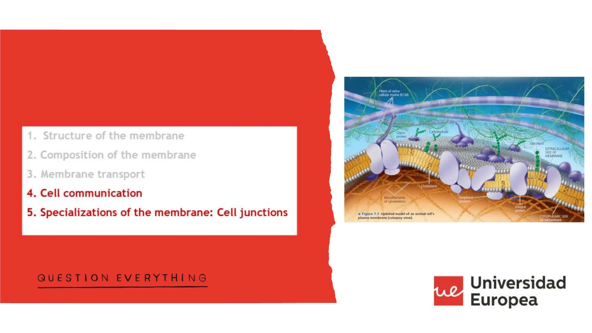
Fibers of extra- celludat matriz (ECM)
1
Carbohydrate Glyco- protein
EXTRACELLULAR SIDE CE MEMBRANE
Cholesterol
Maoflament of cytoskeleton
Parpheril proteins
İndegral protein
A Figure 7.3 Updated model of an animal cell's plasma membrane (cutaway view).
CYTOPLASMIC SIDE OF MEMBRANE
QUESTION EVERYTHING
ul Universidad Europeaue
Membrana Plasmática: Comunicación
Cell communication
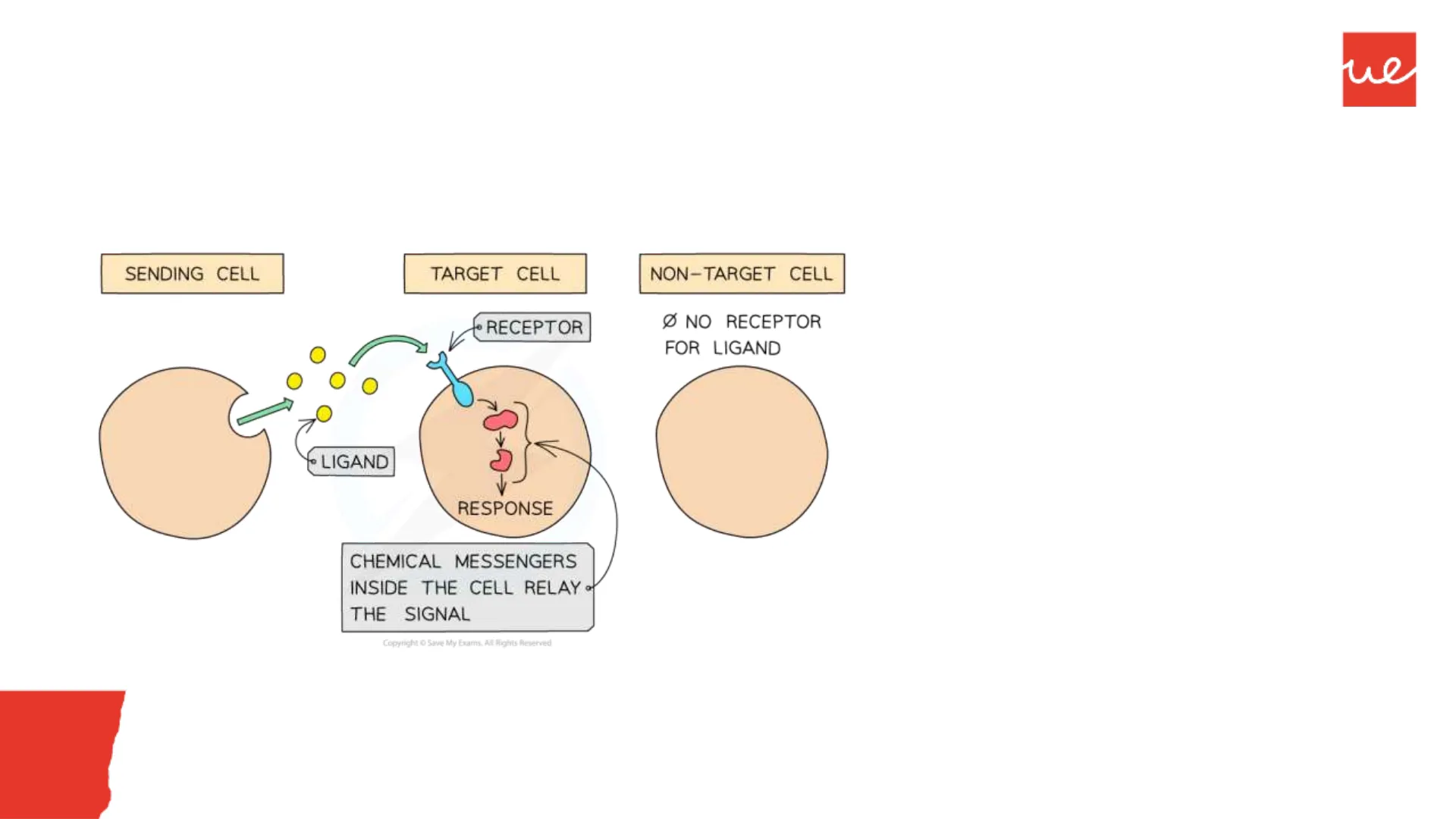
The signaling cell produces a signaling molecule that is detected by the target cell.
SENDING CELL
TARGET CELL
NON-TARGET CELL
RECEPTOR
NO RECEPTOR FOR LIGAND
LIGAND
RESPONSE
CHEMICAL MESSENGERS INSIDE THE CELL RELAY - THE SIGNAL
Copyright O Save My Exams. All Rights Reserved
In order to detect a signal (that is, to be a target cell), a cell must have the right receptor for that signal.
Signaling molecules are the ligands of those receptors.
- Most animal cells send and receive signals, acting as both signaling cells and target cellsue
Membrana Plasmática: Comunicación
Cell communication: What needs to happen for cells to communicate?
- Stimulus reception: A message molecule is recognized by a specific receptor on the membrane or inside the cell.
- Transduction of signal: The signal is transmitted across the membrane to effector molecules on the inner surface or within the cytoplasm.
- Cellular response: Changes in gene expression, enzymatic activity, or ionic concentration.
- End of response by destruction or inactivation of the signal molecule.
Signal Molecule
Receptor
1
Reception
Cytoplasm
Extracellular Fluid
Plasma Membrane
Relay Molecule
2
Transduction
Signal-Transduction Pathway
3
Response
Activation of Cellular Responses
Membrana Plasmática: Comunicación
Importance of cell-to-cell communication
"To regulate their development and tissue organization " To control their growth and division " To coordinate their functions
They communicate in three ways
- They secrete chemical compounds that carry signals to other distant cells
- Through direct contacts between membrane-bound proteins
- GAP junctions: directly communicate the two cytoplasms
bla bla bla bla Ha bla bla bla bla bla bla 2 bla bia bla bla bla bla bla bla bla bla bla bla Ha bla bła bla bla bla bla bla Artiguno
Moreculos de comunicación a Satinca
Señalización por contacto
bla bla bla bla bla bla
Membrana Plasmática: Comunicación
Comunicación Celular
ue
Through: Signal molecules
Through: GAP junctions
Through: Direct contact
Unión tipo gap
Antígeno
Célula presentadora
Conexón
Moléculas de comunicación intercelular a distancia
Célula T helper
Señalización por contacto
Membrana Plasmática: Comunicación
Comunicación Celular
ue
Through: Signal molecules
Through: GAP junctions
Through: Direct contact
Unión tipo gap
Antígeno
Célula presentadora
Conexón
Moléculas de comunicación intercelular a distancia
Célula T helper
Señalización por contacto
Inside Chemical Signaling
Signaling mechanisms
Autocrine/Paracrine
Receptor
Cells secrete local chemical signals, which are so quickly captured, destroyed, or immobilized that they only act on nearby cells (or the sending cell itself, autocrine)
(Short distance)
Synaptic or neuronal
Neurotransmitter
Neurotransmitter
Neuron
Neuron
Electrical signal
Electrical signal
Target cell
Neuron
Electrical signal
It is a special kind of paracrine signalling but only occurs in the nervous system.
Cells secrete neurotransmitters into the synaptic cleft as a response to an electrical signal.
Then neurotransmitters bind to receptors in the postsynaptic cell.
Endocrine
Endocrine cell
Cell without receptor
Cell with receptor
-Target cell
No response
Response
Specialized endocrine cells secrete hormones, which travel through the bloodstream and act on target cells that are widely distributed throughout the body.
(Long distance)
Neuroendocrine
Neurohormone
Neuron
Cell without receptor
Cell with receptor
No response
Response
Same mechanism as in the endocrine system, but hormones are secreted by neurons.
Hormone
Membrana Plasmática: Comunicación
EXTRACELLULAR SIGNAL MOLECULE
1
2
O
0
C
O
O
0
-
O
O
0
Active cell-surface receptor
INTRACELLULAR SIGNALING
Transduction
TARGET PROTEINS
metabolic enzyme
gene regulatory protein
cytoskeletal protein
CELLULAR RESPONSE
altered metabolism
altered gene expression
altered cell shape or movement
76
Nucleus
Modification of gene expression, development
- The same signal can induce different responses in different target cells.
5
Responding cell
8
Signal transduction proteins and second messengers
6
Modification of cellular metabolism, function, movement
7a
Effector protein
What happens once the signal molecule reaches its target cell?
RECEPTOR PROTEIN
Signaling cell
3
9
INTRACELLULAR SIGNALING PROTEINS
Inactive cell-surface receptor
Membrana Plasmática: Comunicación
ue
According to the chemical nature of the signal molecules
- Hydrophilic
- Hydrophobic
According to the type of signalling molecule
- Neurotransmitters
- Hormones
- Growth factors
- Pheromones
Receptor in cytosol
Lipophilic signal molecules
-
Receptor in nucleus
Lipophobic or lipophilic signal molecule
Receptor on surface of cell membrane
Membrana Plasmática: Comunicación
ue
Chemical nature of signal molecules
Depending on the chemical nature of the signal molecule, the mechanism of action is different
Hydrophilic/polar
Hydrophobic/nonpolar
External environment
Cytoplasm
Signal transduction pathway
Cellular response
Membrane receptor
Hydrophilic ligand
Signal transduction pathway
Cellular response
Intracellular receptor
Hydrophobic ligand
Plasma membrane
Membrana Plasmática: Comunicación
ue
Chemical nature of signal molecules: Hydrophobic/nonpolar
- They are transported associated with proteins in plasma
- They have cytoplasmic or nuclear receptors on the target cell
(B)
INTRACELLULAR RECEPTORS
small hydrophobic signal molecule
target cell
carrier protein
nucleus
intracellular receptor protein
Mechanism of action of hydrophobic molecules
1. Binding of the signal molecule to its receptor in the cytoplasm or nucleus. 2. Receptor-ligand enters the nucleus of the target cell and interacts with the DNA 3. Induces the expression of specific genes 4. Synthesis of specific proteins
i.e. Steroid hormones
Adosterone
CHOH
Cortinal
CHOH
0
HSC
HO
HC
0
Estradiol
Pregnencione
DO
HIG
HC
HO
Testosterone
OH
HC
HC
Lipid-soluble hormone
Plasma membrane
1
Nuclear membrane
Ribosome
2
Nuclear receptor
3
Hormone-receptor complex
mRNA
5
Hormone- response element
mRNA synthesis
6
4
Proteins produced
mRNA
Nuclear pore
OH
Progesterone
CH
Membrana Plasmática: Comunicación
ue
Chemical nature of signal molecules: Hydrophilic/polar
- They travel in the bloodstream dissolved in plasma
- Their receptors are in the plasma mebrane of the target cells.
(A)
CELL-SURFACE RECEPTORS
plasma membrane
cell-surface receptor protein
hydrophilic signal molecule
target cell
Membrana Plasmática: Comunicación
ue
Mechanism of action of hydrophilic molecules
1. The signal molecule arrives and binds to the specific receptor 2. A signaling cascade occurs that amplifies the signal through second messengers 3. Cellular response occurs
2203
Intracellular signaling cascade through second messengers.
-One signal molecule
G-protein coupled receptor
Adenylyl cyclase
ATP
G protein
- CAMP
Protein kinase A
Phosphorylated- protein
Cell response
Extracellular fluid
Receptor ligand complex
L
Membrane
R
Intracellular fluid
One ligand is amplified into many intracellular molecules
Membrana Plasmática: Comunicación
ue
Mechanism of action of hydrophilic molecules: Ligand-gated channels
The arrival of the signal molecule opens the channel, which allows the passage of specific ions (Ca2+, Cl- , Na+)
1 Acetylcholine binds to two of the five AChR subunits, causing the channel to change shape and open.
Outside of cell
Na+
Acetylcholine (ACh)
Plasma membrane
000
2 The channel is lined with negatively charged amino acids, allowing Na+ to flow into the cell.
9
Acetylcholine receptor (AChR)
Inside of cell
3 Na+ buildup in cells leads to muscle contraction.
LIFE 8e, Figure 15.5
LIFE THE SCIENCE OF BIOLOGY, Eighth Edition @ 2007 Sinaver Associates, Inc. and W. H. Freeman & Co.
Mechanism of action of hydrophilic molecules
Types of receptors (summary)
señal
Receptor
Exterior celular
Membrana plasmática
Citoplasma
Ligand-gated ion channels
The arrival of the signal molecule opens the channel, which allows the flow of ions (Ca2+, Cl-, Na+)
G-protein-coupled receptors
The arrival of the ligand causes the receptor to interact with an intracellular protein called protein G, which activates a signaling cascade.
Receptors with enzymatic activity
The binding of the ligand to the receptor modifies the enzymatic activity (tyrosine kinase, tyrosine phosphatase ... ) of another area of the protein on the cytoplasmic or inner side of the cell, causing an intracellular signaling cascade
Membrana Plasmática: Comunicación
ue
Mechanism of action of hydrophilic molecules: G protein-coupled receptors
(A)
(B)
(C)
1 Hormone binding to the receptor activates the G protein. GTP replaces GDP.
2 Part of the activated G protein activates an effector protein that causes changes in cell function.
Outside of cell
Signal (hormone)
Activated effector protein
/ GDP
GTP
GDP
G protein- linked receptor
nactive G protein
Inactive effector protein
Activated G protein
The GTP on the G protein is hydrolyzed to GDP.
Cellular responses
Inside of cell
When a G-protein-coupled receptor binds its extracellular signal molecule, the activated receptor signals to a G protein on the opposite side of the plasma membrane, which then turns on (or off) an enzyme (or an ion channel; not shown) in the same membrane.
G protein coupled receptors
Mechanism of action of hydrophilic molecules
G protein-coupled receptors
Ligand Receptor G protein Enzyme
(1st messenger)
2nd messenger
Ligand" (1st messenger) binds to the receptor. The receptor changes shape and activates.
The activated receptor binds to a G protein and activates it.
The G protein changes shape (turns "on"), causing it to release GDP and bind GTP (an energy source).
Activated G protein activates (or inactivates) an effector protein by causing its shape to change.
Extracellular fluid
Effector protein (e.g., an enzyme
Ligand
Receptor
GTP
C
Activated effector enzymes catalyze reactions that produce 2nd messengers in the cell.
Common 2nd messengers include cyclic AMP and Ca2+.)
GTP
G protein
GTP
GDP
Inactive 2nd- messenger
Active 2nd messenger
Second messengers activate other enzymes or ion channels. Cyclic AMP typically activates protein kinase enzymes.
Activated Kinase enzymes
388
- Ligands include hormones and neurotransmitters.
Cascade of cellular Responses
(The amplification effect is tremendous. Each enzyme catalyzes hundreds of reactions.)
Kinase enzymes activate other enzymes. Kinase enzymes transfer phosphate groups from ATP to specific proteins and activate a series of other enzymes that trigger various metabolic and structural changes in the cell.
Intracellular fluid
Second messengers:
ue
They are intracellular molecules that participate in the transmission of the signal to the target molecule.
Signal amplification occurs
Signaling cascade
L
Cellular response
Ca2+
Common 2nd messengers:
AMPc
GMPc
IP 3
Can’t find what you’re looking for?
Explore more topics in the Algor library or create your own materials with AI.
