Lecture: Membrane Biophysics, Midwestern University
Slides from Midwestern University about Lecture #2: Membrane Biophysics. The Pdf explores cellular membrane biophysics, detailing lipid and protein composition, transport mechanisms like simple and facilitated diffusion, and active transport. It covers ion channels, aquaporins, and solute carriers, suitable for university-level Biology students.
See more12 Pages

Unlock the full PDF for free
Sign up to get full access to the document and start transforming it with AI.
Preview
Learning Objectives
- Be able to describe the lipid and protein composition of neuronal cell membranes and relate the composition to the entry of polar or non-polar substances into cells.
- Be able to explain the terms: diffusion, facilitated diffusion, mediated transport, and active transport.
- Be able to describe different types of transporters in the cell membrane and their properties.
The Cell
Cell Membrane
The cell membrane separates the cell from its environment. The general functions of the cell membrane include:
- Physical isolation. The cell membrane is a physical barrier separating the cell's intracellular fluid from the surrounding extracellular fluid.
- Regulation of exchange with the environment. The cell membrane controls the entry of ions and nutrients into the cell, eliminates cellular waste, and releases material from the cell.
- Communication between the cell and its environment. The cell membrane contains proteins that recognize and respond to molecules or changes in its external environment.
- Structural support. Proteins in the cell membrane hold the cytoskeleton, the cell's interior structural scaffolding, to maintain cell shape and secrete substances into extracellular space to create specialized junctions between neighboring cells.
- All biological membranes consist of a combination of lipids and proteins plus a small number of carbohydrates. The ratio of protein to lipid varies widely. Generally, the more metabolically active a membrane is, the more proteins it contains.
Lecture #2 - Membrane Biophysics p. 2-1o
Lipids in Cell Membranes
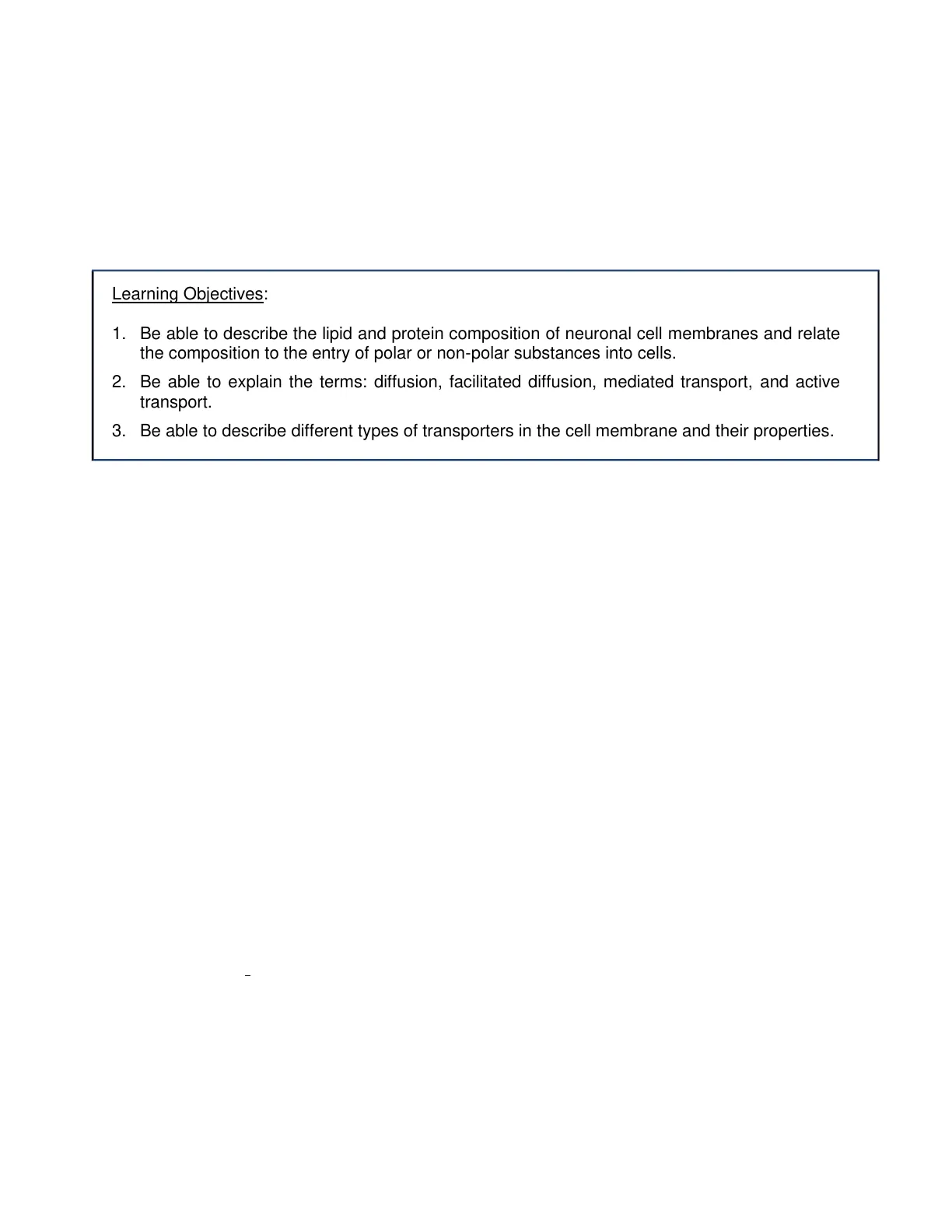
Most of the lipids in biological membranes are phospholipids, which are organized in bilayers with the phosphate heads on the membrane surfaces and the lipids hidden in the center of the membrane. Some membranes also have significant amounts of sphingolipids that are slightly longer than phospholipids. Cholesterol is also a significant part of many cell membranes, is mostly hydrophobic, and inserts itself between the hydrophilic heads of phospholipids. Cholesterol keeps membranes flexible over a wide range of temperatures (Figures 1, 2).
Peripheral proteins can be removed without disrupting the integrity of the membrane. Glycoprotein Transmembrane proteins cross the lipid bilayer. Phospholipid heads face the aqueous intracellular and extracellular compartments. Lipid-anchored proteins Lipid tails form the interior layer of the membrane. Peripheral protein Cytoplasm Cytoskeleton proteins Cholesterol molecules insert themselves into the lipid layer. Fig. 1 Arrangement of the proteins and lipids in a cell membrane.
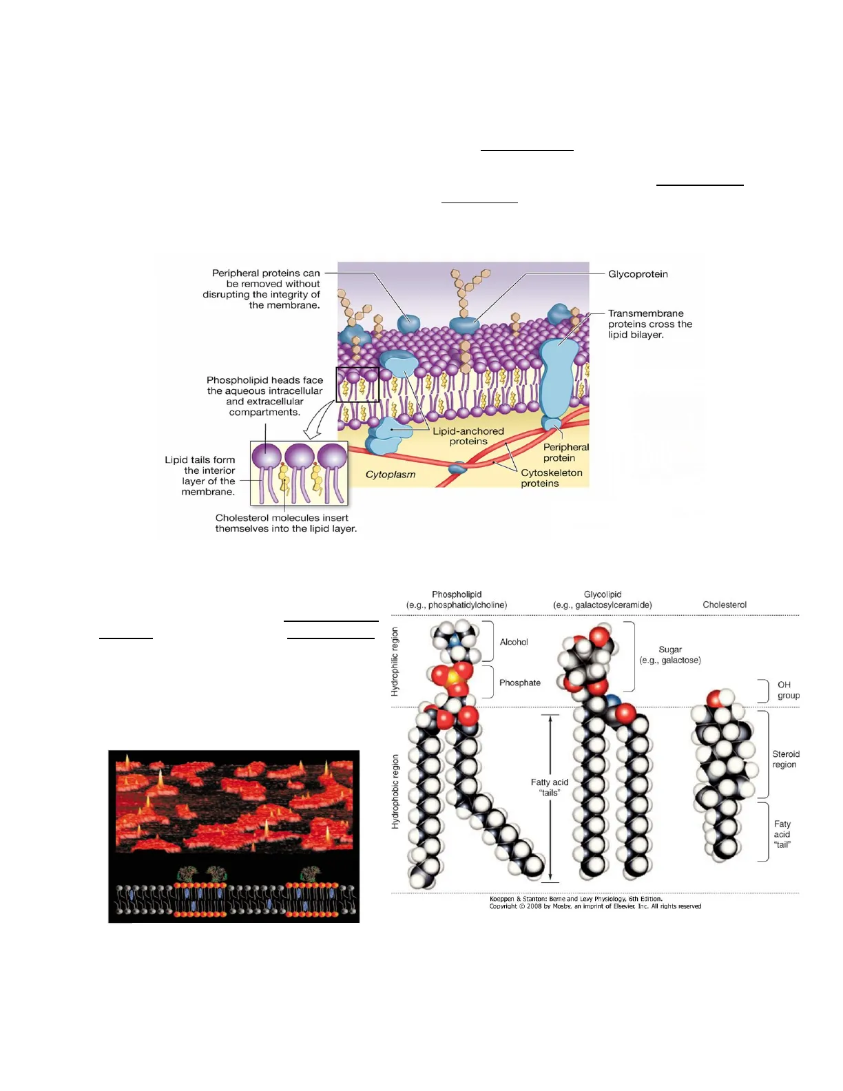
Lipid rafts: specialized patches of the membrane composed of lipid-anchored proteins associated with sphingolipids. Lipid rafts are essential for biological functions such as membrane protein trafficking and signal transduction (Fig. 4). The cell membrane is studded with protein molecules, and the extracellular surface has glycoproteins and glycolipids. Fig. 3 Lipid rafts.
Phospholipid (e.g., phosphatidylcholine) Glycolipid (e.g., galactosylceramide) Cholesterol Hydrophilic region Alcohol Sugar (e.g., galactose) Phosphate OH group Hydrophobic region Steroid region Fatty acid "tails" Faty acid "tail" + Koeppen & Stanton: Berne and Levy Physiology, 6th Edition. Copyright @ 2008 by Mosby, an imprint of Elsevier, Inc. All rights reserved Fig. 2 Models of the major classes of plasma membrane Lecture #2 - Membrane Biophysics p. 2-2o
Membrane Proteins
Membrane proteins can be classified as:
Integral proteins are tightly bound to the membrane, and the only way they can be removed is by disrupting the membrane structure with detergents i.e., transmembrane proteins and lipid- anchored proteins. Peripheral proteins attach to other membrane proteins by noncovalent interactions and can be separated from the membrane by chemical methods that don't disrupt the structure of the membrane. Examples: some enzymes.
Membrane Carbohydrates
o Carbohydrates Most membrane carbohydrates are sugars attached either to membrane proteins (glycoproteins) or to membrane lipids (glycolipids). They are found exclusively on the external surface of the cell where they form a protective layer known as glycocalyx. Surface glycoproteins play a role in the body's immune response, i.e. The ABO blood groups are determined by the number and composition of sugars attached to membrane sphingolipids.
Transport Across Membranes
II. Transport The distribution of solutes in the body depends on whether a substance can cross cell membranes. Water, on the other hand, can move freely in and out of the cells through water-filled channels or a specific type of channel called aquaporin (AQP). We can categorize the movement of molecules across membranes according to their energy requirements (Fig. 4).
A. Passive transport does not require the input of energy other than the potential energy stored in a concentration gradient. B. Active transport requires the input of energy from some outside source, such as the high- energy phosphate bond of ATP.
TRANSPORT ACROSS MEMBRANES Active Passive Vesicular transport (ATP) Protein-Mediated Simple diffusion (concentration gradient) Direct or primary active transport (ATP) Facilitated diffusion (concentration gradient) Exocytosis Ion channel (electrochemical gradient) Endocytosis Indirect or secondary active transport (concentration gradient created by ATP) Phagocytosis Aquaporin channel (osmosis) Fig. 4 Transport across membranes. The movement of substances across membranes can be classified by the energy requirements of transport (in parentheses) or by the physical pathway (through the membrane layer, through a membrane protein, or in a vesicle). Lecture #2 - Membrane Biophysics p. 2-3A.
Passive Transport: Simple Diffusion
Passive transport: Simple Diffusion Diffusion is a passive movement of uncharged molecules down their concentration gradient due to random molecular movement. Diffusion uses only the kinetic energy possessed by all molecules. The net movement of molecules occurs until the concentration is equal everywhere. Once molecules have distributed themselves evenly, the system reaches equilibrium and diffusion stops. Individual molecules are still moving at equilibrium. Note that molecules also move from an area of low concentration to an area of high concentration. The net movement is the difference in movement in these two opposite directions. The dynamic equilibrium state in diffusion means that the concentration has equalized throughout the system, but molecules continue to move. The diffusion of glucose between two compartments of equal volume separated by a permeable barrier is shown in Figure 5.
1 2 2 2 Time A Time B Time C 20 Glucose concentration (mmol/l) Compartment 1 10 - Compartment 2 0 A B C Time Fig. 5 Diffusion of glucose between two compartments of equal volume separated by a permeable barrier. Initially, at time A, compartment 1 contains glucose at a concentration of 20 mM, and no glucose is present in compartment 2. At time B, some glucose molecules have moved into compartment 2, and some are moving at random back into compartment 1. The lengths of the arrows represent the magnitude of the one-way movement. At time C, diffusion equilibrium has been reached, the concentration of glucose is equal in the two compartments (10 mM) and net movement is zero. C1 is the concentration of glucose in compartment 1, C2 is the concentration in compartment 2.
O2 CO2 SMALL, NONPOLAR MOLECULES N2 steroid hormones SMALL, UNCHARGED POLAR MOLECULES H2O ethanol glycerol LARGER UNCHARGED POLAR MOLECULES amino acids glucose nucleosides P IONS H+, Na+ K+, Ca2+ CIT, Mg2+ HCO 3 artificial lipid bilayer Fig. 6 The rate at which a solute crosses a protein-free, artificial lipid bilayer by simple diffusion depends on its size and solubility. Many of the organic molecules that a cell uses as nutrients (red) are too large and polar to pass efficiently through an artificial lipid bilayer that does not contain the appropriate membrane transport proteins.
Diffusion properties : -Passive process -From high concentration to low concentration (chemical gradient) -Net movement until the concentration is equal (equilibrium) -Rapid over short distances -Directly related to temperature -Inversely related to molecular weight and size Lecture #2 - Membrane Biophysics p. 2-4
Passive Transport: Ion Channel
1 1B. Passive transport: Ion Channel In the body, only lipophilic (dissolved in fats) molecules can cross the membrane by simple diffusion. Most of the body molecules are either electrically charged or lipophobic and cannot cross the cell membrane by simple diffusion. They need the help of membrane proteins, in a process called mediated transport. Note that mediated transport can be passive or active. One example of passive transport is the channel. Channels provide corridors for charged molecules to pass through the cell membrane.
One protein subunit of channel Channel through center of membrane protein Channel through center of membrane protein (viewed from above) Fig. 7 Many channels are made of multiple protein subunits that assemble in the membrane. Hydrophilic amino acids in the protein line the channel, creating a water-filled passage that allows ions and water to pass through. Ion channels are found in all cells and are especially important in excitable cells such as neurons and muscle cells. Ion channels are classified by their selectivity, conductance, and mechanism of channel gating (opening and closing). Selectivity is defined as the nature of the ions that pass through the channel. Ion channels can be highly selective, in that they allow only a specific ion through, or non-selective, allowing all or a group of cations or anions through. Conductance refers to the number of ions that pass through the channel. Factors that control gating include membrane voltage, extracellular agonists or antagonists, intracellular messengers (Ca2+, ATP ... ), and mechanical stretch of the plasma membrane.
Water Channels (Aquaporins)
c. Water channels In the case of water molecules, we have specific channels called aquaporin (AQP) channels. The aquaporins are a family of membrane water channels and they are the main routes for water movement in and out of the cell. They are widely distributed throughout the body (brain, lungs, kidneys, salivary glands, gastrointestinal tract, and liver). Cells express different AQP isoforms. Regulation of the amount of water that can enter or leave the cell via AQPs occurs primarily by altering the number of AQPs in the membrane. AQP channels play a role in a variety of physiological processes, including the regulation of exocrine fluid secretion, water, and salt homeostasis, and epidermal hydration.
plasma membrane aquaporins Fig. 8 Shaped like an hourglass, each aquaporin channel forms a pore across the bilayer, allowing the selective passage of water molecules. Shown here is an aquaporin tetramer, the biologically active form of the protein.
Passive Transport: Solute Carriers
D. Passive transport: Solute carriers Solute carriers are a large group of membrane transporters. They bind specific substances that are too polar or too big to cross the membrane and transport them across the cell membrane.
- This type of transport is facilitated diffusion
Fig. 9 Solute carrier. Ions skab Lecture #2 - Membrane Biophysics p. 2-5
Can’t find what you’re looking for?
Explore more topics in the Algor library or create your own materials with AI.
