Circulatory System: Cardiovascular and Lymphatic Systems
Slides about Circulatory System. The Pdf details the heart's anatomy, chambers, valves, and blood vessels, enriched with clear diagrams. The Pdf is suitable for high school Biology students, providing definitions and detailed descriptions of the circulatory system.
See more31 Pages

Unlock the full PDF for free
Sign up to get full access to the document and start transforming it with AI.
Preview
CIRCULATORY SYSTEM
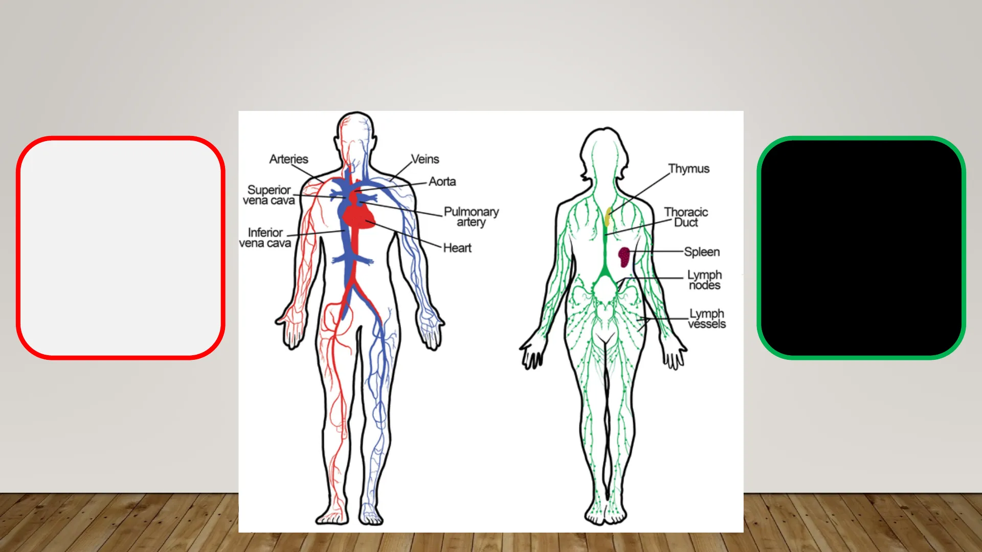
The circulatory system consists of the cardiovascular system and the lymphatic system.
Arteries Veins Aorta Superior vena cava Pulmonary artery Inferior vena cava Heart Spleen Lymph nodes Lymph vessels Blood
- : Heart
: Blood vessels (arteries, veins & capillaries) Thymus Thoracic Duct
The cardiovascular system
The lymphatic system Lymph : Lymph nodes : Lymph vessels (veins and capillaries)
THE CIRCULATORY SYSTEM
The circulatory system distributes nutrients, hormones and oxygen to cells. It also collects the waste products produced during cell metabolism and takes them to the excretory organs responsible for eliminating them.
Digestive system Nutrients Carbon dioxide Respiratory system Endrocrine system (Glands) Hormones Circulatory system distribution and collection Waste products Excretory system Respiratory system Oxygen
THE CARDIOVASCULAR SYSTEM FUNCTIONS
- It transports nutrients from the digestive system to all the body's tissues.
- It takes oxygen from the respiratory system to the cells, and takes carbon dioxide away from them.
- It collects waste products produced by cell metabolism and transports them to the excretory organs responsible for eliminating them.
- It takes specialist defence cells and molecules to tissues infected by pathogenic microorganisms.
- It distributes hormones produced by the endocrine glands, taking them to the cells where they perform their function.
- It helps to regulate body temperature, redistributing heat.
THE CARDIOVASCULAR SYSTEM COMPONENTS
- Heart: A pump that circulates the fluid (blood).
- Blood: A circulating fluid containing the substances and cells that need to be transported.
- Blood vessels: A network of vessels (arteries, veins and capillaries) through which the blood circulates.
Heart Blood Blood vessels
BLOOD COMPOSITION
Blood is made up of: o Blood cells
- Blood plasma (90% water, nutrients, hormones, ions and metabolic waste)
Why is blood red?
D red blood cells blood vessel white blood cell plasma platelets @ 2014 Encyclopædia Britannica, Inc.
BLOOD COMPONENTS
Plasma Salts/Minerals Ko Water Proteins 55% Buffy Coat White blood cells Platelets Centrifugation 1% 45% Red Blood Cells Cleveland Clinic @2021
Blood plasma (55%)
Blood plasma is a yellowish fluid composed mainly of water, but also mineral salts, nutrients, metabolic wastes and hormones.
BLOOD CELLS
Plasma Salts/Minerals Water Proteins 55% Buffy Coat White blood cells Platelets Centrifugation 1% 45% Red Blood Cells Cleveland Clinic @2021
White blood cells (leukocytes)
Blood cells with nucleus. All white blood cells have a defensive function by producing antibodies (lymphocytes) or phagocytosing foreign particles.
BLOOD PLATELETS
Plasma Salts/Minerals Water Proteins 55% Buffy Coat White blood cells Platelets Centrifugation 1% 45% Red Blood Cells Cleveland Clinic @2021
Platelets
They are fragments of cells. They play an essential role in blood clotting.
RED BLOOD CELLS
Plasma Salts/Minerals Water Proteins 55% Buffy Coat White blood cells Platelets Centrifugation 1% 45% Red Blood Cells Cleveland Clinic @2021
Red blood cells (erythrocytes)
They are flat, biconcave cells without a nucleus. They are red because they contain haemoglobin, a protein containing iron that transports oxygen to cells.
Include this draw in your notebook D
BLOOD VESSELS
There are three types of blood vessel with different structures and functions: arteries, capillaries and veins.
Capillary + Arteriole Venules Vein Artery + + + + + +
BLOOD VESSELS: ARTERIES
Arteries
- Function: Arteries take blood from the heart to the rest of the body. Structure: They have thick, elastic walls with a strong smooth muscle layer. They branch into smaller vessels called arterioles.
An artery External connective tissue Endothelium Thick muscle layer ▪ ¡
BLOOD VESSELS: VEINS
Veins
- Function: Veins collect blood from the body and take it to the heart. Structure: They have thinner, less elastic walls than arteries. Veins branch into venules. Flap valves inside the veins let blood flow towards the heart and prevent it from flowing backwards.
An artery External connective tissue Endothelium Thick muscle layer A vein Thin muscle layer Endothelium External connective tissue Flap valves ▪ ¡
BLOOD VESSELS: CAPILLARIES
Capillaries
- Structure: Capillaries are microscopic vessels. Their walls are formed by a single layer of cells called the endothelium.
- Function: They allow the exchange of substances between circulatory system an other systems.
An artery A vein External connective tissue Thin muscle layer Endothelium Endothelium Thick muscle layer External connective tissue A capillary very small lumen Endothelium Sections through the three types of blood vessels
BLOOD VESSELS: TISSUE LAYERS
Endothelium Smooth muscle tissue VENA Connective tissue ARTERIA
THE LAYERS OF THE HEART
The heart is a hollow, muscular organ. It is located between the lungs.
- Endocardium: Epithelial tissue that lines the inside of the heart.
- Myocardium: Middle thickest layer made up of cardiac muscle tissue (involuntary, fast and powerful contractions).
- Pericardium: Double membrane of connective tissue that surrounds the heart.
HEART WALL Pericardium Myocardium Endocardium Parallel Coaching
Include this draw in your notebook 0
STRUCTURE OF THE HEART
SUPERIOR VENA CAVA AORTA PULMONARY ARTERY PULMONARY VEINS LEFT ATRIUM RIGHT ATRIUM MITRAL VALVE PULMONARY VALVE AORTIC VALVE LEFT VENTRICLE TRICUSPID VALVE RIGHT VENTRICLE INFERIOR VENA CAVA The heart is made up of chambers, valves and blood vessels.
STRUCTURE OF THE HEART: CHAMBERS AND VALVES
CHAMBERS
Atria: Two upper chambers with thin walls. Ventricles: Two lower chambers with much thicker walls than the atria. The left ventricle wall is thicker than the right ventricle wall.
Arterial valves + + + + + Left atrium Right atrium Tricuspid valve Mitral valve Right ventricle Left ventricle Interventricular septum +
VALVES
The atria are connected to the ventricles by the Atrioventricular valves: · Tricuspid valve (right side). · Mitral valve (left side). The valves which prevent blood flowing backwards from the arteries into the ventricles are the Arterial or semilunar valves. The heart is made up of chambers, valves and blood vessels.
0 Where are they and What is the function of the coronary arteries and veins?
STRUCTURE OF THE HEART: BLOOD VESSELS
VEINS
- Two vena cavae: superior vena cava and inferior vena cava enter the right atrium.
- Four pulmonary veins: two from the right lung and two from the left enter the left atrium.
Superior vena cava Aorta Pulmonary Artery Pulmonary Veins Pulmonary Veins + + + +
Inferior vena cava
ARTERIES
" The pulmonary artery: leaves the right ventricle and takes blood to the lungs. · The aorta: takes blood from the left ventricle to the rest of the body.
THE CARDIAC CYCLE
1. Atrial systole
The atria, which are full of blood, contract and push blood into the ventricles via the atrioventricular valves (mitral and tricuspid).
2 1 3
3. Diastole
The cardiac muscle relaxes and the atria fill with blood. A new cycle begins.
2. Ventricular systole
1 The atrioventricular valves close to stop blood flowing backwards and ventricles contract_and push blood through the semilunar valves into the aorta and pulmonary artery. The semilunar valves then close 2 Systole: Contraction Diastole: Relaxation
THE HEARTBEAT
120 Semilunar valves close 100 - Semilunar valves open Pressure (mm Hg) 80 Aortic pressure 60 - 40 - Ventricular pressure AV valves open AV valves close 20 - Atrial pressure 0 1st 2nd 3rd Heart sounds 1 2 Prolonged and deep sound. Shorter sound Heartbeats are rhythmic contractions and it indicates that the heart is working. When one of the heart valves does not close properly, other sounds can be heard. These are called heart murmurs.
Aorta Superior vena cava Normal cardiac rythm 70-100 beats per minute Inferior vena cava Pulmonary arteries Pulmonary veins Left atrium Right atrium Left ventricle Right ventricle
HUMAN BLOOD CIRCULATION
Closed because blood always circulates through the blood vessels and never leaves them. Double because blood must flow through the heart twice to complete a circuit. Complete because oxygenated and deoxygenated blood never mix. They circulate through different circuits.
BLOOD CIRCULATION PATHWAY
1. Make a diagram representing the path that a red blood cell will take to provide oxygen to the neurons of the brain, starting from the right atrium and including the names of the chambers, valves, and blood vessels in the correct order. 2. Compare your description with that of your partner and correct and complete your own if necessary. 3. Transform this scheme into a text using linking words.
Capillary bed of lungs where gas exchange occurs Pulmonary veins Pulmonary arteries Pulmonary circuit Aorta and branches Vena cavae Left atrium Left ventricle Right atrium Right ventricle Systemic arteries Systemic veins Oxygen poor, CO2 - rich blood Systemic circuit . Oxygen rich, CO2 - poor blood Capillary bed of all body tissues where gas exchange occurs
BLOOD TYPES: THE ABO SYSTEM
AB0 system Group A Group B Group AB Group O Red blood cell type A B AB O Antibodies in plasma Anti-B Anti-A None Anti-A and Anti-B Antigens in red blood cell A antigen B antigen A and B antigens None
BLOOD TYPES: RH FACTOR
The Rh factor is based on a group of antigens called Rhesus factors. People who have these factors are Rh positive and people who do not are Rh negative.
RH Blood Group Rh factor Rh Positive Rh Negative
TRANSFUSIONS AND BLOOD GROUPS
For a transfusion to be compatible, it is necessary to check that the recipient's plasma has no antibodies against the antigens of the donor's red blood cells.
- Group O Rh- is the universal donor, as it has no antigens (A, B, or Rh factor).
- Group AB Rh+ is the universal recipient, as it has no antibodies (anti-A, anti-B, or anti-Rh).
WHO CAN YOU DONATE TO?
0- 0+ A- A+ B- B+ AB- AB+
WHO CAN YOU RECEIVE FROM?
0- 0+ A- A+ B- B+ V AB- AB+
0 How does the lymph move?
THE LYMPHATIC SYSTEM
Lymph: colourless liquid that circulates through the lymph vessels. It is made up of interstitial fluid, blood plasma and white blood cells. It can also contain microorganisms. Lymph vessels: narrow tubes with closed sacs at the end. Lymph nodes: organs formed by an internal network of fibres and connected by the lymph vessels. Location: Mainly in the groin, armpits and neck. When they are developed are called lymph organs. + + + + + + +
Can’t find what you’re looking for?
Explore more topics in the Algor library or create your own materials with AI.
