Blood and the Circulatory System in Biology for High School
Slides about Blood and the Circulatory System. The Pdf, a presentation for High school Biology, explores the human circulatory system, blood composition, heart function, and coronary artery diseases, with clear titles and illustrations.
See more37 Pages

Unlock the full PDF for free
Sign up to get full access to the document and start transforming it with AI.
Preview
Blood and the Circulatory System
Composition of Blood
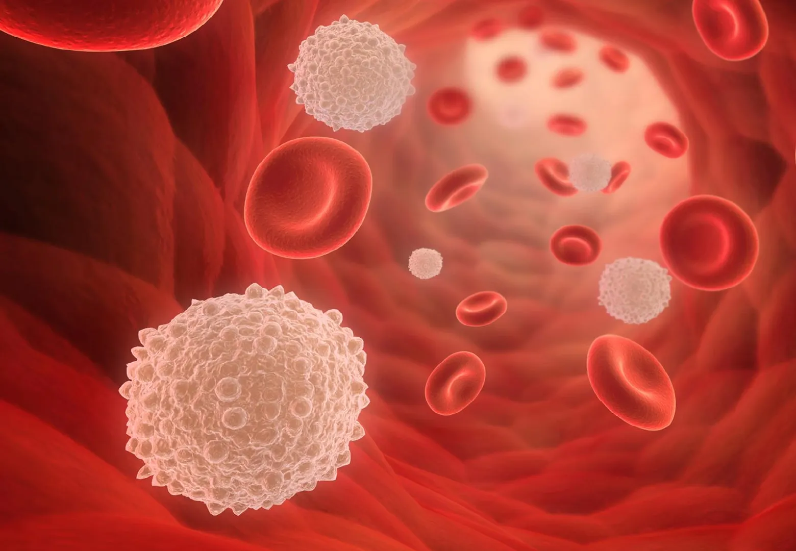
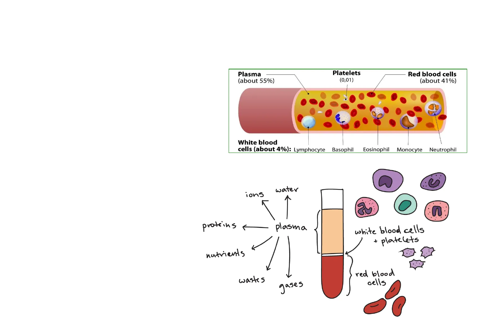
Lesson 1 : Blood and the circulatory systemThe composition of blood: . It is a mixture of cells, solutes and a liquid. · Plasma. . Red Blood Cells. · White Blood Cells.
Plasma (about 55%) Platelets (0,01) Red blood cells (about 41%) White blood cells (about 4%): Lymphocyte Basophil Eosinophil Monocyte Neutrophil water ions n proteins plasma white blood cells + platelets - nutrients wastes red blood cells gasesThe composition of blood: -Plasma Straw-coloured liquid in which all the cells are suspended. Also carries dissolved substances such as glucose, carbon dioxide and urea. USE [ chonpoid Conor ABO/Rh. Patient ABOM ARM BA Stan-Plasma
Blood Plasma Transport
Table 9.2 summarises some of the substances that are transported in blood plasma.
Component Source Destination Notes Water Absorbed in small intestine and colon. All cells. Excess is removed by the kidneys. Various proteins (including fibrinogen and antibodies) Fibrinogen is made in the liver. Antibodies are made by lymphocytes. Remain in the blood. Fibrinogen helps in blood clotting. Antibodies kill invading pathogens. Lipids including cholesterol and fatty acids Absorbed in the ileum. Also derived from fat reserves in the body. To the liver, for breakdown. To adipose tissue, for storage. To respiring cells, as an energy source. Breakdown of fats yields energy - heart muscle depends largely on fatty acids for its energy supply. High cholesterol levels in the blood increase the risk of developing heart disease. Carbohydrates, especially glucose Absorbed in the ileum. Also produced by the breakdown of glycogen in the liver. To all cells, for energy release by respiration. Excess glucose is converted to glycogen and stored in the liver. Excretory substances, e.g. urea Produced by amino acid deamination in the liver. To kidneys for excretion. Most of the urea is removed by the kidneys, dissolved in water to form urine. Mineral ions, e.g. Na+, CI- Absorbed in the ileum and colon. To all cells. Excess ions are excreted by the kidneys. Hormones Secreted into the blood by endocrine glands. To all parts of the body. Hormones only affect their target cells. Hormones are broken down by the liver, and their remains are excreted by the kidneys. Dissolved gases, e.g. carbon dioxide Carbon dioxide is released by all cells as a waste product of respiration. To the lungs for excretion. Most carbon dioxide is carried as hydrogencarbonate ions (HCO3 -) in the blood plasma.
Red Blood Cells
Red Blood Cells Called Erythrocytes. They are the most abundant. Contain haemoglobin a protein that binds with oxygen and transport it around the body. Red blood cells do not have a nucleus Red blood cells, white blood cells and platelets are formed in bone marrow.
White Blood Cells
White Blood Cells Part of the immune system There are different types of white blood cells: Phagocytes: destroy pathogens by phagocytosis. Lymphocytes: produce antibodies, which prevent the spread of microorganisms.
white blood cell red blood cells plasma platelets 1 A phagocyte moves towards a group of bacteria, and flows around them. 2 The phagocyte's cell membrane fuses together, enclosing the bacteria in a vacuole. 3 Enzymes are secreted into the vacuole and digest the bacteria. 4 Soluble substances diffuse from the vacuole into the phagocyte's cytoplasm.
Platelets and Blood Clotting
Platelets Fragments of cells that are involved in blood clotting. When there is a broken blood vessel, platelets: · Prevent infection (stops pathogens getting into the body). · Prevent blood loss. Blood plasma contains a soluble protein called fibrinogen. When a blood vessel is broken, the platelets release a substance that makes the fibrinogen change.
Platelets and blood clotting Blood plasma contains a soluble protein called fibrinogen. When a blood vessel is broken, the platelets release a substance that makes the fibrinogen change. The soluble fibrinogen becomes an insoluble protein called fibrin. Fibrin form fibres which pile up forming a mesh-like structure that helps to seal the wound. The platelets stick together to form clumps. The fibres trapped red blood cells and platelets forming a blood clot. Blood vessels are damaged and the blood contacts new surfaces. A chain reaction occurs which activates blood clotting factors. Platelets become activated. fibrinogen (soluble) fibrin (insoluble) enzyme-catalysed reaction Insoluble fibrin forms fibres which trap blood cells. Platelets stick together and to surfaces. A blood clot is formed.
Blood Groups
Blood groups The AB0 system Two types of protein, A and B, can be present in red blood cell membranes. They are used to classify blood into 4 groups: Group A > People with only protein A Group B > People with only protein B Group AB -> People with protein A and B Group 0 > People with neither protein A or B The Rh system A protein known as the Rh factor can also be present in the blood. Positive (+) > People with Rh protein. Negative (-) > People without Rh protein.
Blood Donors and Recipients
Blood donors and recipients If proteins from one person are introduce into another, it can cause rejection. The blood group of the donor and recipients need to be consider before blood transfusions.
TYPE YOU CAN GIVE BLOOD TO YOU CAN RECEIVE BLOOD FROM A+ A+, AB+ A+, A-, 0+, 0- 0+ 0+, A+, B+, AB+ 0+, 0- B+ B+, AB+ B+, B-, 0+,0- AB+ AB+ EVERYONE A- A+, A-, AB+, AB- A-, 0- 0- EVERYONE 0- B- B+, B-, AB+, AB- B-, 0- AB- AB+, AB- AB-, A-, B-, 0-
Blood Vessels
Blood vessels: artery capillary network vein Figure 9.13: Arteries divide to form capillaries, which join up again to form veins.
Arteries
ARTERIES Blood flows away from heart in arteries. Narrow tube Thick layer of elastic and muscle fibres The heart squirts high pressure blood into arteries with each beat. The walls are thick to withstand the sudden increase in pressure. A wave of stretching then passes along the walls- you feel this as your pulse. After stretching, muscle and elastic fibres in the artery walls contract making the blood flow much more smoothly.
Veins
VEINS Blood returns to the heart in veins. . vein valve Wide tube (facilitates the flow of the low pressure blood) Thin, flexible wall (the low pressure does not require thick vessels to withstand it) Valves to stop blood from flowing backwards. Blood flows under low pressure in veins and so they only need thin walls. As you move, muscles in your skeleton help to push blood along the veins.
Capillaries
CAPILLARIES Arteries divide into narrow capillaries. Capillaries deliver oxygen and nutrients to cells and to take away waste materials. Narrow tube Capillaries gradually join up to form veins. Wall is only one cell thick (facilitates diffusion) Cell wall is very thin to allow faster diffusion of substances into and out of the capillary. Capillaries form a fine network running through the tissues.
Blood Vessel Structure
An artery thick outer wall small lumen thick layer of muscles and elastic fibres smooth lining A capillary very small lumen wall made of a single layer of cells A vein fairly thin outer wall thin layer of muscles and elastic fibres large lumen O smooth lining Figure 9.12: Sections through an artery, capillary and vein. The drawings are not to scale - in reality, capillaries are much smaller than either arteries or veins.
Blood vessels:
Blood vessel Function Structure of wall Width of lumen How structure fits function arteries carry blood away from the heart thick and strong, containing muscle and elastic tissue relatively narrow; it varies with heartbeat, because the walls can stretch and recoil strength and elasticity needed to withstand the high pressure and pulsing of the blood as it is pumped through the arteries by the heart capillaries supply all cells with their requirements, and take away waste products very thin, only one cell thick very narrow, just wide enough for a red blood cell to pass through no need for strong walls, as most of the blood pressure has been lost; thin walls and narrow lumen bring blood into close contact with body tissues veins return blood to the heart quite thin, containing far less muscle and elastic tissue than arteries wide; contains valves no need for strong walls, as most of the blood pressure has been lost; wide lumen offers less resistance to blood flow; valves prevent backflow Table 9.1: Structure and functions of arteries, capillaries and veins.
Main Blood Vessels
Main blood vessels in the human body: vein from arm artery to arm vena cava from head aorta pulmonary veins pulmonary artery vena cava from body hepatic artery hepatic vein renal artery hepatic portal vein artery to small intestine renal vein vein from leg artery to leg
Circulatory Systems
The circulatory systems The blood in the left-hand side of the heart has come from the lungs. It contains oxygen and it is called oxygenated blood. The oxygenated blood is sent around the body, some oxygen is taken by cells for respiration and the blood becomes deoxygenated. The deoxygenated blood returns to the right-hand side of the heart and it is sent to the lungs where it becomes oxygenated. Oxygen diffuses into the I blood from the lungs. Pulmonary system Deoxygenated blood is carried to be lungs. Oxygenated blood is carried from the lungs. Deoxyger ated blood is returned to the right side of the heart. A Oxygenated blond is carried to all the cells in the body from the left side of the heart. right side of heart left side of heart Systemic system Double circulatory system Oxygen diffuses from the bloed to the body cells.
Double Circulatory System
The circulatory systems Double circulatory systems are found in mammals, birds and reptiles. Fish have a circulatory system in which the blood passes only once through the heart to complete a full cycle. Advantage of a double circulatory system: · Helps to maintain a higher blood pressure, meaning that the blood travels slower. · Nutrients and oxygen are delivered more efficiently. 1 Oxygen diffuses into the blood from the gills. heart Oxygen diffuses from the blood to the body cells. Single circulatory system
The Heart
The heart to head to lungs aorta pulmonary artery to body from head pulmonary vein from lungs vena cava left atrium le-way valve right atrium one-way valve le-way valve tendon supporting valve left ventricle vena cava septum from body right ventricle
Can’t find what you’re looking for?
Explore more topics in the Algor library or create your own materials with AI.
