Elisa Method: Understanding Immunoassay Techniques
Slides from University about Elisa Method. The Pdf, a teaching material for Biology students at University, explains the ELISA and Lateral Flow Immunoassay methods, detailing their procedures, principles, and applications, including standard curve construction and comparisons of formats.
See more9 Pages

Unlock the full PDF for free
Sign up to get full access to the document and start transforming it with AI.
Preview
ELISA Method
Non-Competitive ELISA (Sandwich Immunoassay)
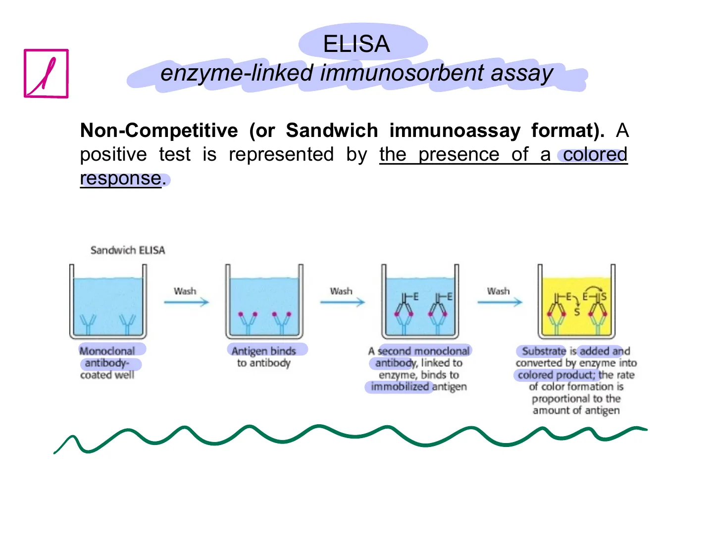
ELISA enzyme-linked immunosorbent assay Non-Competitive (or Sandwich immunoassay format). A positive test is represented by the presence of a colored response. Sandwich ELISAs, use two antibodies specific to the antigen to capture or "sandwich" antigens in the well for detection.
Sandwich ELISA Steps
SANDWICH ELISA STEPS: Wash Wash Wash HE IFE -E EHIS S Monoclonal antibody- coated well Antigen binds to antibody A second monoclonal antibody, linked to enzyme, binds to immobilized antigen Substrate is added and converted by enzyme into colored product; the rate of color formation is proportional to the amount of antigenELISA enzyme-linked immunosorbent assay
Standard Curve Construction
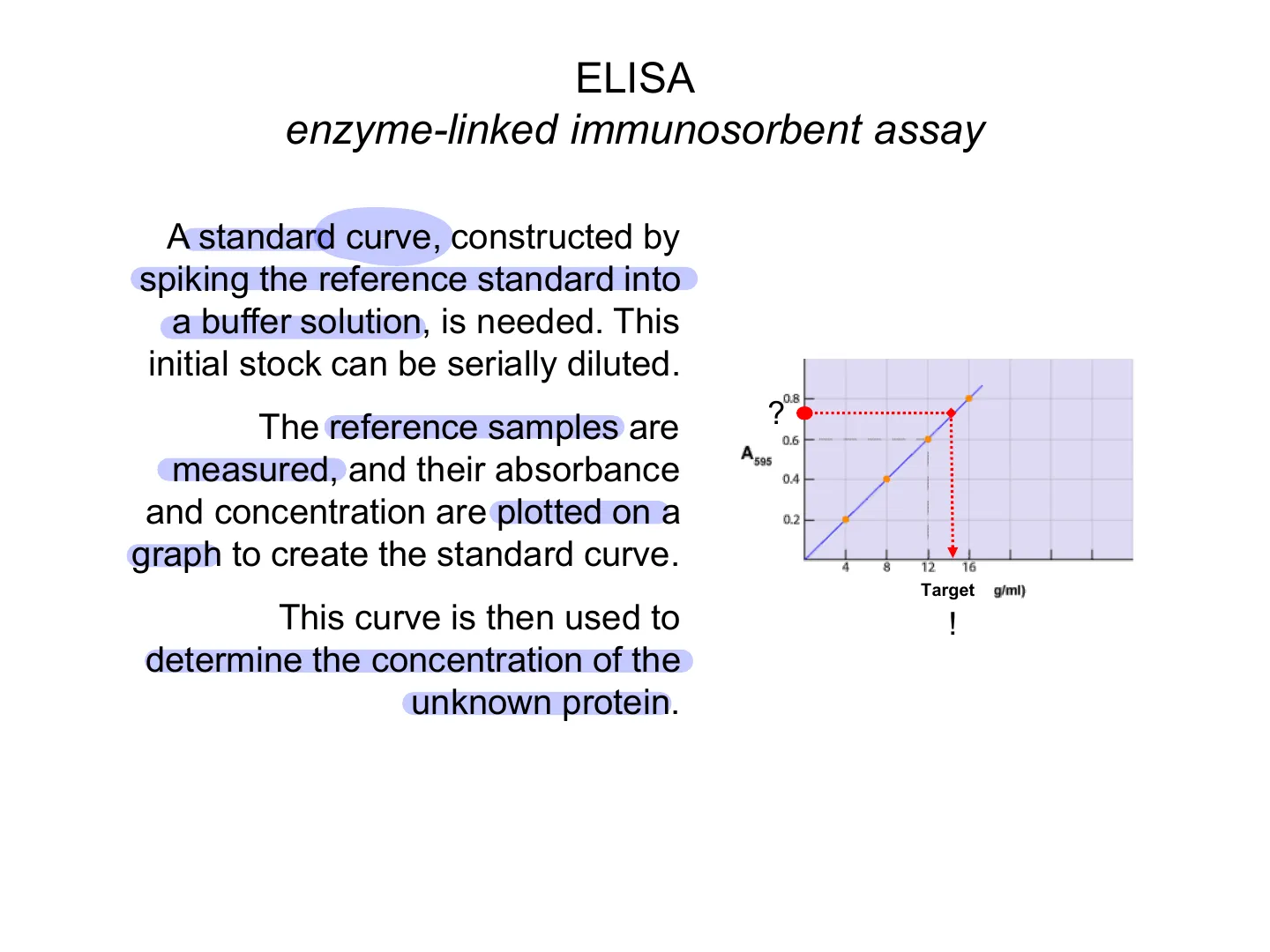
A standard curve, constructed by spiking the reference standard into a buffer solution, is needed. This initial stock can be serially diluted. The reference samples are measured, and their absorbance and concentration are plotted on a graph to create the standard curve. This curve is then used to determine the concentration of the unknown protein.
0.8 ? 0.6 A 595 0.4 0.2 4 8 12 16 Target g/ml) !ELISA enzyme-linked immunosorbent assay
Sandwich vs. Competitive Immunoassay
Sandwich viral spike protein VS. Competitive host IgM/IgG protein immunoassay format
Lateral Flow Immunoassay Method
A lateral flow immunoassay is a simple to use diagnostic device used to confirm the presence or absence of a target analyte, such as pathogens or biomarkers in blood or bodily fluids, or contaminants in water supplies or foodstuffs. The most commonly known type of lateral flow rapid test strip is the pregnancy test.
Lateral Flow Immunoassay Components
2 Reaction Site Test Site Control Site Free antibody (trapstarget) + dye enzyme Fixed antibody (traps target) + dye substrate Fixed antibody (traps free antibody) + dye substrate YY 1 1 Reaction Site Test Site Control Site Rapid test (searching for viral spike protein, lateral flow assay with commercial antibodies)
Non-Competitive Lateral Flow Immunoassay
Non-Competitive (or Sandwich immunoassay format) - used for large molecular weight analytes with multiple antigenic sites. A positive test is represented by the presence of a coloured line. A negative test is represented by the absence of a line.
Positive Result in Non-Competitive Lateral Flow
Reaction Site Test Site Control Site YỶ 1 I Two lines = positive result
Negative Result in Non-Competitive Lateral Flow
Non-Competitive (or Sandwich immunoassay format) - used for large molecular weight analytes with multiple antigenic sites. A positive test is represented by the absence of a coloured line. A negative test is represented by the absence of a line. Reaction Site Test Site Control Site No No YY 1 One line = negative result
Positive vs Negative Control
Positive Control
Positive control gives a response to the experiment. What is a Positive Control (Ctrl+) 10 Ctrl+ A positive control is an experimental control that gives a positive result at the end of an experiment. This type of test always gives the result as a "yes". It is a good indication to know if the test works. Hence, positive controls are used to evaluate the validity of a test. The positive control is not exposed to the experimental test; it is done parallel to it. The positive control is used to get the expected result. This positive result ensures the success of the test. If the positive control does not give the expected result, the experiment must be repeated.
Negative Control
Negative control does not give any response. What is a Negative Control 10 Ctrl- A negative control is an experimental control that does not give a response to a test. The negative control is a not exposed to the experimental test directly. It is done parallel to the experiment as a control experiment. The negative control is used to confirm that there is no response to a reagent (or any other parameter) used in the test. In order to get a good result from a negative control, one should ensure that there is no net response to the test. Hence, negative controls are helpful in identifying outside influences on the experiment.
SARS-COV-2 Diagnostics Example
SARS-CoV-2 Components
SARS-COV-2 DIAGNOSTICS EXAMPLE: Spike Glycoprotein (S) M-Protein Hemagglutinin-esterase dimer (HE) Envelope RNA and N protein E-Protein
SARS-CoV-2 Diagnostic Methods
SARS-CoV-2 diagnostics: - Molecular (nucleic acid, RT-qPCR; probe - PCR primers) - Rapid (viral spike protein, lateral flow test = commercial antibodies) - Serological (host IgM/IgG protein, ELISA, lateral flow test = commercial antibodies)
Can’t find what you’re looking for?
Explore more topics in the Algor library or create your own materials with AI.
