Lectures on Graded Potentials and Action Potentials, Midwestern University
Slides from Midwestern University about Graded Potentials and Action Potentials. The Pdf explores the mechanisms of depolarization, repolarization, and hyperpolarization, along with the characteristics and differences between the two types of potentials in Biology for University students.
See more20 Pages

Unlock the full PDF for free
Sign up to get full access to the document and start transforming it with AI.
Preview
Learning Objectives
- Be able to define depolarization, repolarization, and hyperpolarization.
- Be able to list the characteristics of graded potentials and action potentials. Be able to explain how they differ.
- Be able to explain the concepts of decremental conduction and summation.
- Be able to explain the mechanisms for the generation of a variety of graded potentials: i.e., excitatory postsynaptic potentials (EPSPs), inhibitory postsynaptic potentials (IPSPs), and end- plate potentials (EPPs)
- Be able to explain the concept of the action potential threshold and the mechanisms responsible.
- Be able to explain the mechanisms for absolute and relative refractory periods.
- Be able to explain the ionic mechanisms responsible for the different phases of the action potential.
- Explain the mechanisms for non-decremental conduction via local current flow and saltatory conduction.
- Be able to explain how the Na-K ATPase pump contributes to the resting membrane potential both directly and indirectly.
- Be able to list the factors which determine the conduction velocity of neurons.
Introduction to Membrane Potential Changes
The membrane potential can change over time, allowing signals to be transmitted. These variations in membrane potential are mediated by specific ion channels opening and closing, affecting the membrane's conductance to the ions. Excitable cells (e.g., neurons) use two types of changes in membrane potential (graded potentials and action potentials) to generate and transmit signals (information). There are two basic types of electrical signals:
- Graded Potentials (also known as generator or receptor potential) -They have variable strength -They are used for short-distance communications
Lecture #4&5- Graded and Action Potentials p. 4/5-1· Action Potentials -They are very brief, have large depolarizations -They are rapid over long distances
Graded Potentials
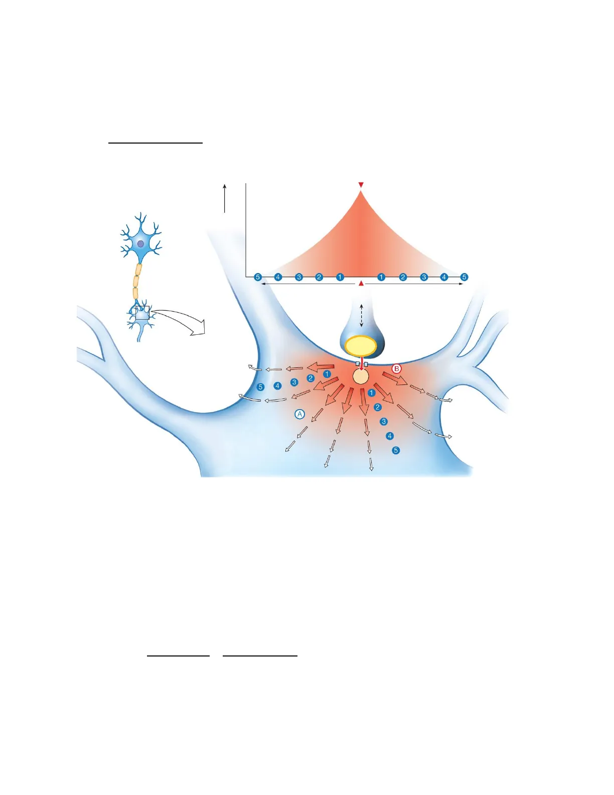
Graded potentials dissipate with distance due to current leak out of the cell
Graded potential amplitude (strength) mV 5 4 3 2 1 1 2 3 4 5 Distance Stimulus Distance point of origin Axon terminal Stimulus Postsynaptic neuron B 1 + 2 Na 5 4 1 2 A 3 1 Current leak out of the cell 4 5
Fig. 2. Graded potentials. their magnitude varies directly with the strength of the stimulus. Q. At which point of the neuron will the graded potential be stronger, A or B?
Characteristics of Graded Potentials
- Graded potentials are signals of variable strength
- They are used for short-distance communication
- The graded potential can initiate an action potential if the depolarizing graded potential is strong enough when it reaches the trigger zone within a neuron
- Can be depolarizing or hyperpolarizing
- Occur in the dendrites and cell body, and less frequently, near the axon terminal
- Travel passively, uniformly in all directions
Lecture #4&5- Graded and Action Potentials p. 4/5-2 3- don't require voltage-gated channels decay with distance from the site of stimulation
Channels Involved in Graded Potentials
Channels involved in graded potentials: Ion channels responsible for graded potentials are: -ligand-gated (extracellular ligands such as neurotransmitters) -mechanosensitive, or temperature-sensitive channels
Factors Affecting Graded Potential Strength
Why do graded potentials lose strength as they move through the cell? Because two factors play a role:
- Current leak. In this case, the membrane of the neuron cell body has open leak channels that allow positive charge to leak out into the ECF. Some positive ions leak out of the cell across the membrane decreasing the strength of the depolarization wave as it travels through the cell.
- Cytoplasm resistance. The cytoplasm provides resistance to the flow of electricity.
Events of a Graded Potential
- An external stimulation causes gated ion channels to open. The right stimulus activates ion channels that are mechanically, chemically, or light-gated.
- A small area of the membrane is hyperpolarized or depolarized.
- If the stimulus opens Na+ channels, Na+ moving into the cell causes the membrane potential to become more positive, resulting in depolarization; the inside of the membrane becomes more positive, and the outside becomes more negative.
- If the stimulus opens K+ channels, K+ moving out causes the membrane potential to become even more negative, resulting in hyperpolarization. This type of graded potential can prevent the generation of an action potential (it has an inhibitory effect). It causes the membrane to drop below-resting membrane potential so that it's impossible to get to the threshold potential to generate an action potential.
- The magnitude of a voltage change is proportional to the strength of the stimulus. The stronger the stimulus, the more gated ion channels that open and the greater the depolarization or hyperpolarization.
Types of Graded Potentials
- Receptor potentials occur in specialized sensory receptor cells
- result from transduction processes (the conversion of an energy stimulus into an electrical potential). This can be due to the opening of either mechano-, thermo-, or ligand-gated channels.
Lecture #4&5- Graded and Action Potentials p. 4/5-3A) B) C) Channel closed Channel open Mechanical force Channel closed Blade Channel Open Heat Ion flux through open pore Ion flux through open pore
Fig. 3. A) Model for the ligand-gating channel. B) The TRPV1 channel responds to heat as well as to chemical signals such as capsaicin. This channel consists of four subunits, which form a central cation-selective pore. Heat is thought to open the TRPV1 channel pore by displacing membrane lipids closely associated with the channel, leading to a structural rearrangement that opens channel gates (red). C) Piezo, an example of a mechanosensitive channel, has a unique structure that includes three subunits and large blades on the extracellular side of the channel. The Piezo channel pore is thought to open when mechanical force displaces the blades, leading to the rearrangement of the structure of the rest of the channel.
- Postsynaptic potentials occur in neurons:
- Result from the activation of ligand-gated channels.
- Depolarizing potentials are called excitatory postsynaptic potentials (EPSPs); hyperpolarizing potentials are called inhibitory postsynaptic potentials (IPSPs). -Excitatory Postsynaptic Potentials (EPSPs) - These synaptic potentials increase the likelihood of a postsynaptic action potential occurring. The postsynaptic response to an excitatory neurotransmitter opens mixed (non-specific) cation channels which carry both Na+ and K+. This results in depolarization. Why? It is considered excitatory since it brings the membrane closer to the threshold for firing action potentials. -Inhibitory Postsynaptic Potentials (IPSPs) - These synaptic potentials decrease the likelihood of a postsynaptic action potential occurring. The postsynaptic response to inhibitory neurotransmitters opens CI- or K+ channels. This causes hyperpolarization. It is considered inhibitory since it furthers the cell from the threshold.
Fig. 4. Excitatory postsynaptic potential (EPSP) and inhibitory postsynaptic potential (IPSP). Stimulation of the presynaptic neuron occurs at the time indicated by an arrow. The graded potentials shown are larger than the typical EPSP or IPSP; most individual EPSPs and IPSPs are less than 1 mV in amplitude.
Membrane potential (mV) Threshold -EPSP -70 -70 1 - IPSP 10 20 10 20 Time (ms) Time (ms)
Lecture #4&5- Graded and Action Potentials p. 4/5-4 Ligand(A) (B) Summed EPSPs (Synapses E1 + E2) IPSP Summed EPSP + IPSP (Synapses E1 + I) Summed EPSPs + IPSP (Synapses E1+I+E2) Excitatory E1 +20 1 Postsynaptic membrane potential 1 Excitatory E2 + -20 Cell body -40 Threshold Axon V rest Dendrites -60 E1 or F2 E1 + E2 I E1 + E1 E2 + I Time (ms)
Fig. 5. Summation of postsynaptic potentials. A microelectrode records the postsynaptic potentials produced by the activity of two excitatory synapses (E1 and E2) and an inhibitory synapse (I). (B) Electrical responses to synaptic activation. Stimulating either excitatory synapse (E1 or E2) produces a subthreshold EPSP, whereas stimulating both synapses at the same time (E1 + E2) produces a suprathreshold EPSP that evokes a postsynaptic action potential (shown in blue). Activation of the inhibitory synapse alone (I) results in a hyperpolarizing IPSP. Summing this IPSP (dashed red line) with the EPSP (dashed yellow line) produced by one excitatory synapse (E1 + I) reduces the amplitude of the EPSP (solid orange line) while summing it with the suprathreshold EPSP produced by activating synapses E1 and E2 keep the postsynaptic neuron below threshold so that no action potential is evoked. Purves et al. Neuroscience 5th ed.
- Endplate potentials (EPPs) occur in muscle cells
- As a result of the activation of ligand-gated channels, caused by the opening of mixed cation channels that carry both Na+ and K+. Generally, it is very similar to EPSPs.
Repolarization After a Graded Potential
The restoration of the resting membrane potential (repolarization) following a graded potential is a passive process due to the same forces that originally determined the resting potential, i.e., the permeabilities of the membrane for K+ and Na+ and the driving forces for K+ and Na+.
Action Potentials (AP)
- Are very brief, large depolarizations that travel from a neuron's trigger zone to the end of its axon
- Do not decrease in strength with distance. The conduction is non-decremental; the action potential does not decrease in amplitude over distance.
- The action potential is caused by the activation of the voltage-gated ion channels, most often the Na+ voltage-gated ion channel
- Voltage-gated ion channels in the axon membrane open sequentially as electrical current passes down the axon
- Action potentials are "all-or-none" phenomena because they either occur as a maximal depolarization (if the stimulus reaches a threshold) or do not occur at all (if the stimulus is subthreshold, meaning below the threshold). Therefore, amplitudes are all-or-none. Once a stimulus is of sufficient strength, an explosive, relatively large (~100 mV) response of standard size (the action potential) occurs.
Lecture #4&5- Graded and Action Potentials p. 4/5-5 + EPSP (Synapse E1 or E2) (Synapse I) Record Inhibitory I Postsynaptic membrane potential (mV) 0
Can’t find what you’re looking for?
Explore more topics in the Algor library or create your own materials with AI.
