Drug Development and Formulation Nuclear Magnetic Resonance Spectroscopy
Slides from Www.biosite.dk about Drug Development and Formulation Nuclear Magnetic Resonance Spectroscopy. The Pdf explores NMR spectroscopy, focusing on quantification via 1H NMR, with practical examples using aspirin and paracetamol spectra for quantitative analysis in Chemistry at University level.
See more15 Pages

Unlock the full PDF for free
Sign up to get full access to the document and start transforming it with AI.
Preview
Nuclear Magnetic Resonance Spectroscopy - Lecture 2
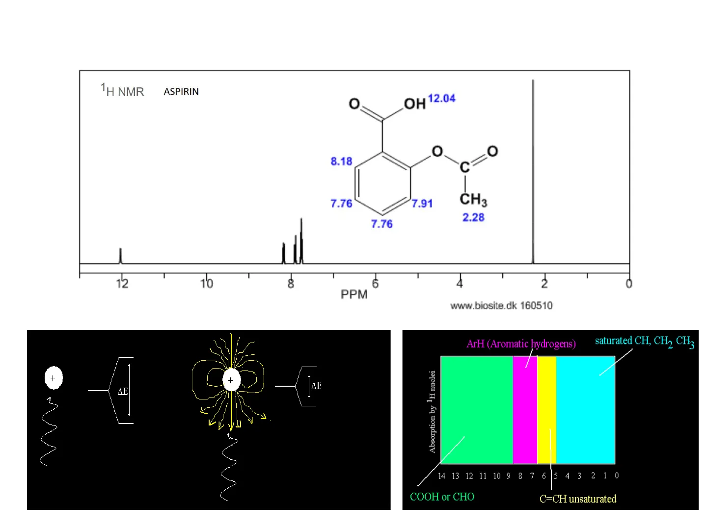
1H NMR Aspirin
O
OH 12.04
O
O
8.18
C
7.76
7.91
CH3
2.28
7.76
12
10
8
6
4
2
PPM
www.biosite.dk 160510
+
ΔΕ
ΔΕ
Absorption by 1H nuclei
14 13 12 11 10 9 8 7 6 5 4 3 2 1 0
COOH or CHO
C=CH unsaturated
saturated CH, CH, CH3
ArH (Aromatic hydrogens)
NMR - n + 1 rule
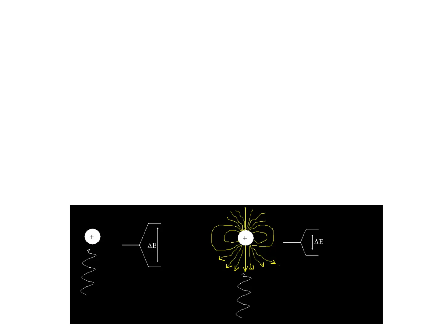
- Remember that each hydrogen has a magnetic moment which at
any one moment in time can be going with the applied field or
against it. - Also remember also how the chemical shift is influenced by the
electron density around a hydrogen i.e. electrons have their own
magnetic moment that always goes against the applied filed,
weaking it and making it "easier" for the hydrogen to "flip".
+
+
ΔΕ
ΔΕ
NMR - n + 1 rule: Impact of Neighbouring Hydrogens
- Electrons are not the only thing that can impact on the strength of the
applied magnetic field. Neighbouring hydrogens are close enough for
their own magnetic moment to impact on the chemical shift of a
hydrogen environment. - Consider the hydrogen environment in blue below. It has one
neighbouring hydrogen atom in red. At any one moment in time the red
neighbouring hydrogen could have a magnetic moment aligning with the
magnetic filed or against it.
エ ー
H
1
CI-C
1
-
C-Br
CI
Br
NMR - n + 1 rule: One Neighbour
- When the neighbouring hydrogen's magnetic moment aligns with the
applied field it strengthens it making it harder for the blue hydrogen to
flip (more energy needed, so downfield shift). - When the When the neighbouring hydrogen's magnetic moment goes
against the applied field it weakens the strength of the applied magnetic
field (a bit like electrons do), so less energy is needed for the blue
hydrogen to flip (so upfield shift).
H
I-
1
I
CI-
C
C-
- Br
I
CI
Br
Neighbouring H going
with the field and "making
it more difficult" for H, so
it's harder to flip
(downfield shift)
Neighbouring H going
against the field and
"helping/shielding" H,
so it's easier to flip
(upfield shift)
Downfield
Chemical shift
Upfield
NMR - n + 1 rule: Two Neighbours
- When you have more that one neighbouring hydrogen atom, both act
individually to either "help" or "hinder" at any one moment of time. - With two neighbours there are three combinations of things that could
happen - 1. Both neighbours go with the applied field
- 2. Both neighbours go against the applied field
- 3. One goes with and the other goes against (statistically speaking this is
twice as likely as either 1 or 2)
H
H
1
H
H
H
HL
H
H1
I
I
I
I
I
CI-C
-C
-
H 1
CI-C-C-
-H CI-C
-
-C:
-
CI
Br
CI
Br
CI
Br
CI
Br
Harder to flip -
so downfield
shift
Easier to flip -
so upfield shift
No change
overall as these
cancel out
No change
overall as these
cancel out
c-
-H
I
-
I
C
-H1 CI-
NMR - n + 1 rule: Two Neighbours and Triplet Splitting
H
H 1
H
H
H
HL
H
H1
I
1
I
I
1
1
CI-C-
C.
-
H1
CI-C-
C
CI-C-
.C
- H 1
CI-C-
-
-c-
-H
I
I
I
I
CI
Br
CI
Br
CI
Br
CI
Br
Harder to flip -
so downfield
shift
Easier to flip -
so upfield shift
No change
overall as these
cancel out
No change
overall as these
cancel out
So our blue H is "split" into three peaks in the ratio of 1:2:1
Triplet
Downfield
Upfield
Chemical shift
This is where the n + 1 rule
comes from. A hydrogen
environments is split into the
number of neighbouring
hydrogen plus one.
0-
NMR - n + 1 rule: Three Neighbours and Quartet Splitting
H
H 1
CI-C
-C-
- H1
I
CI
H
H
H1
H
H1
H
HL
I
I
I
I
CI-C-
-C
-
-
I
CI
H
1
H
H
H
H
1
1
1
I
I
1
CI
一 工
1
CI
HL
CI
HL
H
H
I
1
- H
-
I
CI
H
L
upfield (easier)
Quartet
Downfield
Upfield
Chemical shift
Downfield (Slightly harder)
-
CI
H
-H1
CI-C
-C-H|CI-C
-C
H
-
CI
H 1
H1
CI-C
-C
-H
CI-C-C-H CI-C-
C
- H
I
-
Upfield (slightly easier)
1
CI-C-
-C
1
Downfiled (harder)
1
HL
NMR - n + 1 rule: Pascal's Triangle
No Coupled
Hydrogens
C-C-C-H
1
doublet
1
1
triplet
1
2
1
quartet
1
3
3
1
1
4
6
4
1
J
J
1
5
10
10
5
1
1
6
15
20
15
6
1
J
J
J
H-C-C-H
1
MA Quarte
H
Pascal's Triangle
Three Coupled
Hydrogens
C
C
H
c-C-C-H
1
H
1
MA A Singlet
J
One Coupled
Hydrogen
MA Doublet
Two Coupled
Hydrogens
H-C-C-H
Č
H
1
W A Triplet
Lets try some examples.
Nuclear Magnetic Resonance Spectroscopy Examples
0
H3C-C
B
O-CH2-CH3
C
A
A = 1.26
C = 4.11
TMS
C
B
A
7
6
5
4
3
2
1
0 8 ppm
B = 3.75
エ ー
CI-C-C-C-CI
HHH
BAB
A = 2.20
TMS
N
7
6
5
4
3
2
1
0 8 ppm
B = 2.04
HHH
Examples: Molecule Matching
Which molecule is most likely to match the following NMR
2 2
1
2
1
3
6
6
4
2
0
X
OH
X
N
O
O
NMR Predictor:
http://www.nmrdb.org/new_predictor/index.shtml?v=v2.138.0
I
Quantification in NMR
The examples we have used are very basic, and do not fully represent
the power of NMR or its wide applications. NMR is often used
qualitatively. It is good to be aware of this. There is a video to
accompany this that you should watch in your own time.
Other atoms can be analysed by NMR e.g. 13C. This is not so useful in
Quantification due to its low abundance.
b
I-Z:
f
C
dN
e
b
a
:0:
c
b
0-
200
180
160
140
120
100
80
60
40
20
0
CDS-02-660
ppm
H.
Quantification by 1H NMR
NMR is usually run on pure compounds for qualitative analysis - but
since the size of the peaks in 1H NMR are proportional to the number
of protons giving rise to the signal, it can also be used quantitatively.
O
3H
HO
N
-C-CH3
1H
2H
2H
1H
11
10
9
8
7
6
5
4
3
2
1
0
HSP-43-745
ppm
This is the NMR of
paracetomol. The
integrations just show
the ratio of Hs in each
environment - however
the size of the peaks
also tell you how much
is there.
Quantification by 1H NMR: Internal Standard Method
When using NMR quantitatively you need to select a single peak that does
not overlap other peaks i.e. the CH3 signal at 2ppm.
0
H
HO
-C-CH3
11
10
9
8
7
6
5
4
3 2 1
0
HSP-43-745
ppm
You then select a suitable
"internal standard" that has a
clear signal well away from
the rest of the paracetamol
peaks e.g trioxane which
gives a singlet at 5ppm due to
its 6 equivalent H's.
0
H2(
CH2
0
Ò
H2
s-Trioxane
Make up a solution of the paracetomol tablet in a suitable NMR solvent
(e.g. CDCl3) and add a known concentration of trioxane. The spectrum you
obtain will have all the paracetomol peaks plus an extra peak for trioxane.
You can then use the following equation to calculate the concentration of
paracetamol:
Number of moles
Paracetamol
Intergral for
Paracetamol
Number of protons
giving signal
Number of moles of s-trioxane
s-trioxane intergral
Number of protons
giving signal
Quantification by 1H NMR - Example Calculation
Example:
0
N
6H |f
S-Trioxane
HO
Drug A
e
2H
a
h
1H
3H
b
c
1H
2H
1H
d
1H
1
12
11
10
9 8
7
6
5
3
2
1
0
Chemical shift 8
A tablet (weighing 250 mg) containing Drug A (RMM 207) was
crushed and 200 mg of tablet powder was dissolved in exactly 5
cm3 of CDC1, containing 0.15M s-trioxane. In the resulting
spectrum the integration of the trioxane peak was 45 and the
integration of peak e was 18. Use this information to calculate the
mass of Drug A in the tablet.
Nuclear Magnetic Resonance Spectroscopy Example Solution
Example:
5 cm3 of 0.15 M s-
trioxane = 7.5 x 10⁻⁴
moles
0
f
g
a
N
h
e
6H
|f
S-Trioxane
C
d HO
Drug A
18
b
e
2H
a
45
h
1H
3H
c
00
1H
2H
1H
d
1H
12
11
10
9 8
7
6
5
4
3
2
1
0
9.0 x 10⁻⁴ moles
Chemical shift 8
Number of moles
Drug
18
Intergral for
Drug
Number of protons
giving signal
2
Number of moles of s-trioxane
7.5 x 10⁻⁴ moles
s-trioxane intergral
45
Number of protons
giving signal
6
Since RMM of drug A is 207 then there is 0.1863 g (186.3 mg) in the
200 mg tablet powder analysed, so there is 232.8 mg of Drug A in the
original tablet (250 mg).
Can’t find what you’re looking for?
Explore more topics in the Algor library or create your own materials with AI.
