Vascular System Layout Part 2: detailed anatomy of the venous system
Document from Università Degli Studi Di Milano about Vascular System Layout Part 2. The Pdf describes the detailed anatomy of the venous system, focusing on systemic, head, neck, thoracic, and limb veins, suitable for university-level Biology students.
Ver más21 páginas

Visualiza gratis el PDF completo
Regístrate para acceder al documento completo y transformarlo con la IA.
Vista previa
General Characteristics of Systemic Veins
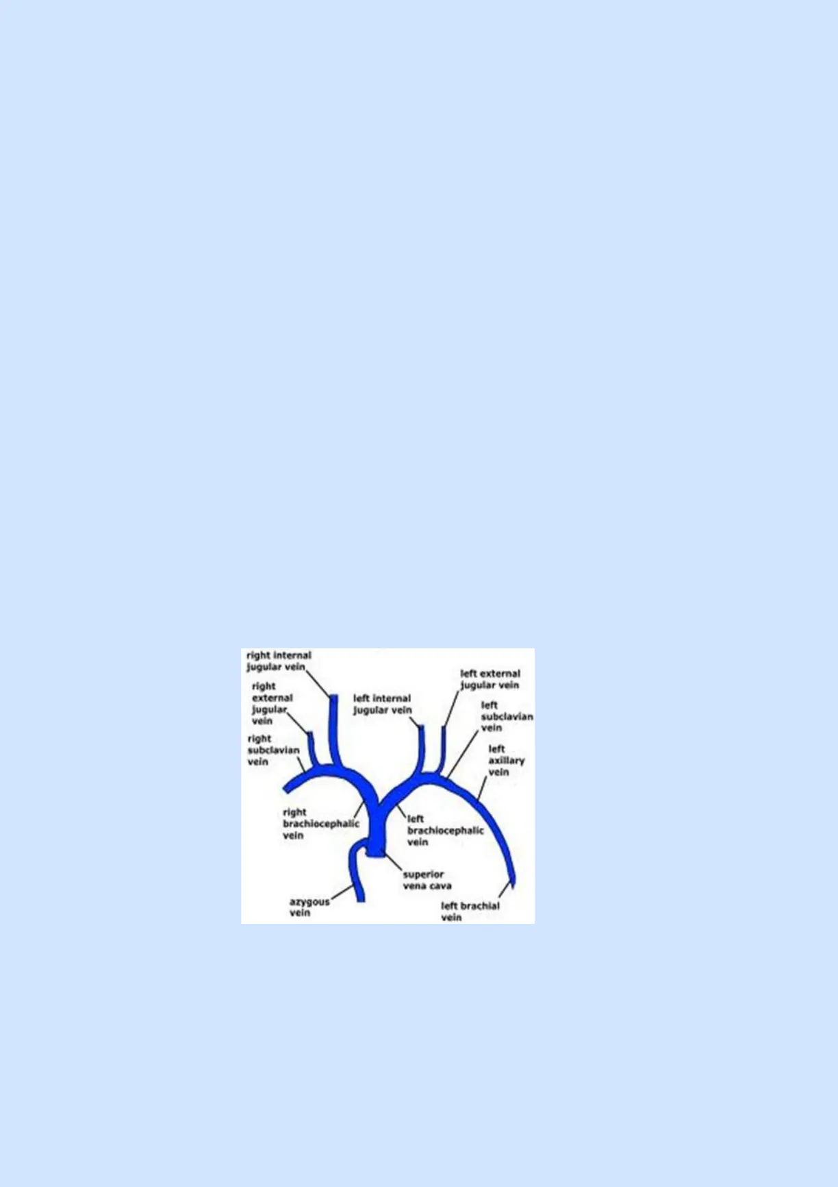
1. General characteristics of systemic veins In the systemic circulation, there are three major veins entering the right atrium: · Superior vena cava (SVC) · Inferior vena cava (IVC) · Coronary sinus A feature of veins that is not present in arteries is that veins can be superficial, and they also commonly arrange to form plexuses, which are networks of anastomoses, and parallel veins. The description of veins can begin from the superior vena cava that reaches the right atrium. This vein is formed by two other large veins: the brachiocephalic trunk (or innominate veins). Differently from arteries, in venous organization there is a right-left symmetry, in fact each brachiocephalic trunk is formed by joining two large veins, the internal jugular vein (coming from the head and the intracranial structures) and the subclavian vein (coming from the upper limb and axilla). The subclavian vein is also joined by a smaller vein, the external jugular vein, which drains blood from the extracranial structures. Another important system of veins that joins the superior vena cava is the azygos system, made of three veins: · Azygos vein · Hemiazygos vein · Accessory hemiazygos vein The superior vena cava collects blood from the supradiaphragmatic regions.
right internal jugular vein right external jugular vein left external jugular vein left internal Jugular vein left subclavian vein right subclavian vein left axillary vein right brachiocephalic vein Meft brachiocephalic vein superior vena cava azygous vein left brachial vein
Head and Neck Veins
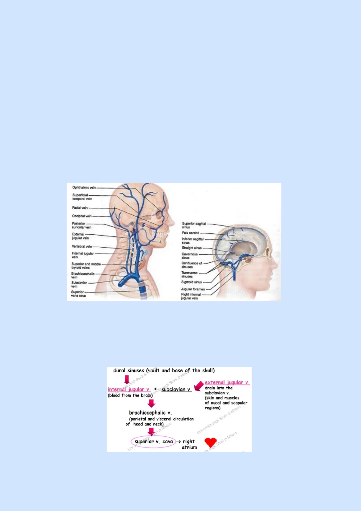
2. Head and neck veinsThe two brachiocephalic veins are posterior to the sternum. The internal jugular veins drain most of the blood from the brain, running from lateral to internal side with respect to the common carotid arteries (extend together with carotid arteries). At the base of the neck, they join the subclavian vein to form the brachiocephalic trunk. The external jugulars drain some of the scalp and the face and they are tributaries of the subclavian. Blood from the brain is drained and transported by very special blood vessels, the dural sinuses, which extend along the meninges enveloping the central nervous system. In particular, part of their wall is made by the dura mater. These sinuses are located superiorly but also at the base of the brain. They are many, and some important ones include: · Superior sagittal sinus. · Transverse sinuses. · Cavernous sinus: dangerous in case of trauma, because it also contains the carotid arteries and some cranial nerves. · Sigmoid sinus: that later joins the internal jugular vein, exiting the brain through the jugular foramen. Ophthalmic vein Superficial temporal vein Facial vein Occipital vein Posterior- auricular voin Superior sagittal sinus Falx cerebri External- jugular vein Inferior sagittal sinus Vertebral voin Straight sinus Internal jugular vein Cavernous sinus Superior and middle thyroid veins Confluence of sinuses Brachiocephalic vein Transverse sinuses Subclavian vein Sigmoid sinus Superior vena cava Jugular foramen Right internal jugular vein To summarise, the head and neck veins begin in the vault and base of the skull with the dural sinuses that join in the internal jugular vein. Meanwhile, the subclavian vein receives blood from the external jugular vein that drains the skin and muscles of nucal and scapular regions. Together, subclavian and internal jugular form the left and right brachiocephalic veins that finally reach the superior vena cava to get to the heart.
dural sinuses (vault and base of the skull) I degli Studi di Milan internal jugular v. + subclavian v. (blood from the brain) external jugular v. drain into the subclavian v. (skin and muscles of nucal and scapular regions) brachiocephalic v. (parietal and visceral circulation of head and neck) Milano Università degli Studi di Milano superior v. cava > right atrium "tà degli Studi di Milano
Veins in the Thorax
3. Veins in the thoraxIn the thorax, there are both visceral and parietal branc" Regarding the visceral veins, an important structure is , gous vein system draining blood from the back, the thoracoabdominal wall and the thoracic viscera. This system is made of three veins: · Azygos vein: leads blood from the right intercostal veins to the superior vena cava. · Hemiazygos vein: collects blood from last five or six left intercostal veins and drains into the azygos vein. · Accessory hemiazygos vein: collects blood from first six or seven left intercostal veins to the azygos vein. The azygos vein system is also joined by two other visceral veins, the bronchial vein and the esophageal vein.
Right superior intercostal vein Left superior intercostal vein Superior vena cava Arch of azygos DE Accessory hemiazygos vein Azygos vein Communication of azygos and hemiazygos veins Intercostal veins Hemiazygos vein
Veins in the Abdomen and Pelvis
4. Veins in the abdomen and pelvis In the abdomen, blood is drained by a large vein, the inferior vena cava, formed by joining of the two common iliac veins and joined by many other veins. A very important one of these is the portal vein, which receives blood from small intestine, spleen and other parts of the abdominal cavity and brings it to the liver, where it is filtered and collected by the hepatic veins that drain into the superior part of the inferior vena cava to get to the heart. Therefore, the inferior vena cava receives blood from the subdiaphragmatic territories of the body. In particular, each of the two common iliac veins that join to form the IVC drains blood from an internal iliac vein (coming from the pelvis wall and other organs of this region) and an external iliac vein (coming from the lower limbs thanks to the connection with the femoral veins). The inferior vena cava also receives some parietal veins (lumbar and the inferior phrenic veins) and some visceral veins, which include: · Renal v. (draining the kidneys) · Adrenal v. · Genital v. · Hepatic v. contain blood filtered by the liver and transported to it by the portal vein.inferior v.cava (subdiaphragmatic territories) Universi internal iliac v. (from pelvis wall and organs) common iliac v. un external iliac v. (from femoral v.) studi · inferior v. cava receives: parietal veins: lumbar v. di di inferior phrenic v. Stu visceral v .: renal v. adrenal v. genital v. hepatic v. Università degli Studi di Moreover, in the abdomen an asymmetry can be noted, because the left gonadal and suprarenal veins drain into the left renal vein (which later goes into IVC), while the right gonadal and suprarenal veins drain directly into IVC (receives 2 renal veins, right gonadal and suprarenal veins).
Inferior phrenic vein Hepatic veins Inferior vena cava Left suprarenal vein Right suprarenal vein Renal veins Left ascending lumbar vein Right gonadal vein Lumbar veins Left gonadal vein L-5 Common iliac vein External iliac vein- Internal iliac vein
Hepatic Portal System
5. Hepatic portal system On the contrary, as above mentioned, blood from organs located in the abdominal cavity is drained by different veins (such as the superior mesenteric and splenic veins) that then join into the hepatic portal vein, which enters the liver through its hilum, branching inside the liver and influencing its microanatomy. The superior mesenteric vein and the splenic vein join together to form the hepatic portal vein. The splenic vein comes from the spleen but also collects blood from the pancreas and from the large intestine. The inferior mesenteric veins also collect blood from the small intestine and drains into the splenic vein. After blood is filtered in the liver, it is collected by the hepatic veins. uni Interior vena cava (not part of hepatic portal system) Hepatic veins Liver Spleen Gastric veins Inferior vens cava Hepatic portal vein Splenic vein ersità deg Right gastroepiploic vein Inferior mesenteric vein ità degli S Superior mesenteric gar Small Intestine hiver Large intestine Rectum Università degli Studi di MThis whole system is known as the hepatic portal system, and together with the portal system of the hypophysis and is an important system. It is composed of two capillary beds in between an artery and a vein, indeed there is an artery distributing blood in organs of the digestive system forming then different capillary beds. The blood is collected by the hepatic portal vein that reaches the liver forming a second capillary bed. Here, the blood interacts with hepatocytes, involved in the processing and storage of its nutrients but also in the detoxification of toxins and drugs. Finally, the filtered blood is collected by the hepatic veins and from here gets to the inferior vena cava.
Arterial blood Venous blood Stomach and intestine Liver Nutrients and toxins absorbed Liver cells (hepatocytes) Nutrients and toxins leave Hepatic portal vein Hepatic vein First capillary bed Second capillary bed (liver sinusoids) Hepatic portal system
Blood Supply of the Limbs
6. Blood supply of the limbs Blood vessels extend in the limbs forming neurovascular bundles that contain the artery, some veins and nerves, all sheeted by connective tissue. In relation to the blood supply of the upper limbs, the most important artery is the subclavian artery, which runs laterally onto the first rib under the clavicle. When it enters the axilla, the artery changes name, becoming axillary artery that continues as brachial artery into the upper arm and then splits into radial and ulnar arteries. These last two finally reach the hand forming its blood supply through palmar arches, dorsal arches and digitals. These arteries extend in the flexor surfaces to avoid collapse and the blocking of blood flow during movement of the different parts of the limb.
Vertebral artery Common carotid arteries Thyrocervical trunk Right subclavian Costocervical trunk artery Suprascapular ariery Left subclavian Thoracoacromial artery Axillary artery Brachiocephalic trunk Subscapular artery Posterior intercostal Anterior circumflex- Anterior humeral artery intercostal artery Brachial artery Internal thoracic Deep artery Lateral thoracic Ulnar collateral arteries Descending aorta Common interosseous artery Radial artery Ulnar artery Università degli Studi Università degli Studi di Milano Deep palmar arch Superficial palmar arch Digitals
Veins in Limbs
7. Veins in limbs In limbs, there are two types of circulation: Deep circulation: includes veins extending together with arteries in neurovascular bundles. · It contains veins, following the corresponding arteries. · Veins share the name of the accompanying artery. · Most deep veins double to one artery. · They extend and transport blood from the same territory. Superficial circulation: no relation with arteries, only veins are present. Deep and superficial circulations are connected by anastomoses (anastomotic tracts), so blood from the superficial circulation can reach the deep one.
artery Posterior circumflex- humeral artery arteries artery of arm artery Inferior vena cava
¿Non has encontrado lo que buscabas?
Explora otros temas en la Algor library o crea directamente tus materiales con la IA.
