Anatomy Lecture Notes: Central Nervous System and Brain Functions
Document from University about Anatomy Lecture Notes Section 3: Nervous System. The Pdf, a set of anatomy notes, describes the central nervous system, focusing on the brain and cerebrospinal fluid, suitable for University Biology students. It includes diagrams and covers topics like hydrocephalus.
Mostra di più18 pagine

Visualizza gratis il Pdf completo
Registrati per accedere all’intero documento e trasformarlo con l’AI.
Anteprima
Nervous System Anatomy
The Central Nervous System: The Brain and the Spinal Cord The nervous system is anatomically and functionally divided into two parts, the Central Nervous System (the brain and the spinal cord) and the Peripheral Nervous System (the ganglia, and 12 pairs of cranial nerves, plus 31 of pairs of spinal nerves). The Peripheral Nervous System (PNS) can be further delineated into the Somatic Nervous System (SNS) which integrates control over skeletal muscle, and the Autonomic Nervous System (ANS) which for the most part automatically regulates vital internal organs and systems.
The Brain
In anatomy, we can divide the brain into six (6) parts in terms of information processing:
- Cerebrum
- Diencephalon
- Midbrain
- Cerebellum
- Pons
- Medulla oblongata
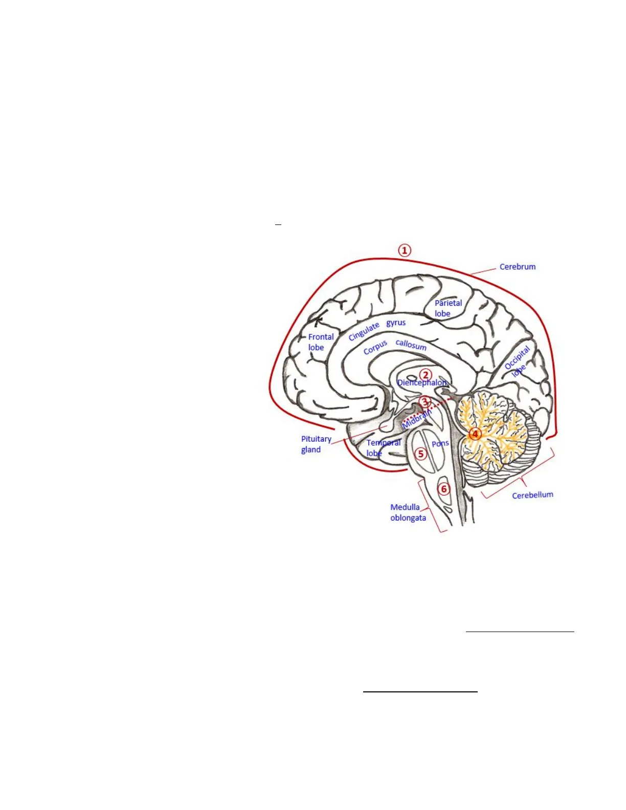
To the right is a mid-sagittal section of the brain showing various regions and the six major divisions (red circled numbers) of the human brain from the highest to lowest level of information processing.
1 Cerebrum Parietal lobe Cingulate gyrus Frontal lobe Corpus callosum Occipital lobe 2 0 Diencephalon 3 Miabrann. Pituitary gland Pons Temperat lobe 5 6 Cerebellum Medulla oblongata
Cerebrum Functions
The cerebrum is the largest most developed area of the human brain (see above) and is considered to be the center of the highest functions. Its major functions include: Awareness of sensory perception; voluntary control of movement (regulation of skeletal muscle movement); language; personality traits; sophisticated mental activities such as thinking, memory, decision making, predictive ability, creativity and self-consciousness.
The cerebrum is composed of 5 lobes, below is some basic information about them:
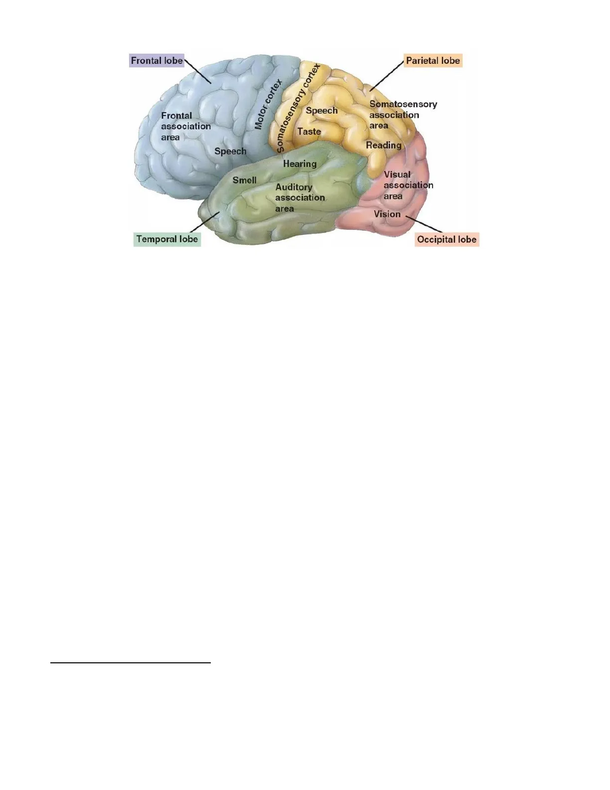
The Frontal Lobe - Encased within the frontal bone is the largest and most complex of the 5 lobes, concerned with higher intellectual functions and behavioral aspects of humans. The primary motor cortex controls the movement of skeletal muscles of the body.
The Parietal Lobe - Protected by the parietal bones of the skull, this lobe is primarily concerned with the interpretation and integration of body sensory inputs. The somatosensory cortex is associated with reception and perception of touch, vibration, temperature, and general body senses. Also involved in spatial orientation, movement coordination, reading, writing and mathematical computation.2
Frontal lobe Parietal lobe Motor cortex Somatosensory cortex Speech Somatosensory association area Frontal association area Taste Reading Speech Hearing Smell Visual association area Auditory association area Vision Temporal lobe Occipital lobe
The Temporal Lobe - Contains the auditory cortex for the perception of sound, and the olfactory cortex for the sense of smell. Also houses the language cortex usually in left hemisphere and participates in recognition and interpretation of language. The amygdala and hippocampus (limbic system) connect to the temporal lobe and aid in memory formation related to emotions, the sense of smell and sound.
The Occipital Lobe - This lobe contains the primary visual cortex for visual information interpretation. The eyes transmit images to the visual cortex, which then determines colors, identify objects, shapes, and other aspects of visual perception. Visual information is sent to the parietal lobes and temporal lobes for further processing. The occipital lobe is also for reading comprehension; depth perception; recognition of object movement. The parietal and temporal lobes use visual info to perform tasks like as opening a door and connecting visuals with retained memories.
The Insula Lobe - A key role is visceral perception or awareness of internal organs and various bodily states. When the bladder is empty we are not consciously aware of it, but when it becomes full, we become aware of it. The gustatory cortex is responsible for taste perception, located in the insula the frontal lobes. The insular lobe also enables us to read our own emotions and be aware of them.
Brain Anatomy Vocabulary
Sulcus (sulci) - grooves on surface of cerebrum. Gyrus (gyri) - fold of brain tissue between sulci. Fissure - deep groove, separating hemispheres.
There are three (3) kinds of cerebral functional area:
- Sensory areas - for receiving incoming sensory information.
- Association areas - for analysis and placing information into context.
- Motor areas - for sending out a response from the cerebrum to lower levels.
Two significant gyri on Cerebrum:
- On Frontal Lobe: Precentral gyrus (motor). This region is the Primary Motor Cortex.
- On Parietal Lobe: Postcentral gyrus (sensory). This region is the Primary Somatosensory Cortex.3
Posterior Motor Sensory Anterior Sensory map in postcentral gyrus Hand Wrist Elbow Arm Shoulder Trunk Knee Leg Hip Neck Head Arm Elbow Hand Fingers Thumb Knee Thumb Eye Neck Brow Toes Lips Face Lips 9211 Jaw Tongue Pharynx Tongue Swallowing Primary motor cortex (precentral gyrus) Primary somato- sensory cortex (postcentral gyrus) S Intra- abdominal
Cerebrum General Anatomy
General Anatomy of Cerebrum - there are 2 Hemispheres - Left and Right.
Left Right 13 10
Left Hemisphere Functions
The Left Hemisphere Controls writing, movement of the right side of body. Usually dominant in language and tasks that involve symbolic reasoning. Language, logic, analytical, sequential, verbal tasks (fragmentary information processing).
Right Hemisphere Functions
The Right Hemisphere Controls touch, movement of the left side of body. Typically superior at non-verbal, visual and spatial tasks. Spatial perception, artistic and musical endeavors (holistic information processing).
Motor map in precentral gyrus Fingers Foot Nose Face Eye Genitals Teeth Gums Jaw Forearm Hip Trunk4
Cerebral Communication: Axon Fibers
Cerebral Communication: The major groups of axon fibers and tracts of the central white matter.
a) Association Fibers - are axons (fibers) connecting different cortical areas on the same side of the cerebrum to each other, that is, within the same hemisphere (L or R). Short association fibers like the arcuate fibers connect adjacent gyri. Long association fibers, like the longitudinal fibers connect distant part of the cerebral cortices within the hemisphere.
b) Commissural Fibers - connect an area in one hemisphere with an area in the opposite hemisphere. The corpus callosum is the largest set of commissural fibers in the brain, having from 200 to 300 million axons crossing from one hemisphere to the other. There are two much smaller but important examples called the anterior commissure and the posterior commissure.
c) Projection Fibers - also known as projection tracts of the brain are a type of white matter tract that connects the cortex with other areas in the CNS, e.g. deep nuclei, brainstem, cerebellum or spine. They may be efferent (motor), which can sometimes be called 'descending' tracts, or afferent tracts (sensory) which can sometimes be called 'ascending' tracts.
Reticular System
The Reticular System - widespread connections, ideal for arousal of the brain as a whole. The Reticular activating system (RAS) - Maintains consciousness and alertness. Functions in sleep and arousal from sleep. This RAS is predominantly located in the Pons and the MO. When aroused, it sends signals form the pons and MO "up" to the higher centers in the brain for conscious awareness.
Limbic System: The Emotional Brain
The Limbic System - The Emotional Brain Still within the cerebrum, the limbic system is a group of structures on the medial aspect of each hemisphere and the diencephalon. It is more of a functional system than an anatomical one. The limbic system is the "emotional brain", participating in the creation of emotional states such as fear, pleasure, anger, affection, arousal, etc. and processing vivid memories associated with those states.
Cingulate Gyrus
The Cingulate Gyrus is an arch-shaped convolution situated just above the corpus callosum, on the medial aspect of the cerebral hemispheres. Involved in information processing for decision making, planning, allows us to shift between thoughts and inter-relate them. It also interprets pain as unpleasant, an integral part of learning and learned avoidance practice.
Hippocampus
The Hippocampus is a complex brain structure embedded deep in the temporal lobe. It is thought to have a major role in learning and memory. It's named after its resemblance to a type of sea-horse. This structure is described as plastic, where neuroplasticity is the ability of the brain to create and reorganize new synaptic connections, especially in response to learning or after experiencing injury, since the hippocampus can become damaged by various stimuli. For example, high, prolonged levels of cortisol (known as the stress hormone) are associated with shrinkage of the hippocampus. Another way to think of this is that worrying too much can shrivel your brain. So worry less.
This area has a role in spatial processing and navigation, integrating spatial relationships and memories. This may explain why some people have a better sense of direction than others, in that their hippocampus is more integrated with their others senses than others. So let them have the map, it's just better for everyone. It also functions in consolidating information and memories during restorative sleep.
Amygdala
The Amygdala takes its name after the almond-shaped nuclei that is nestled within the temporal lobe, it5 is part of a collection of nuclei located deep in the temporal lobe. It appears to have a primary role in detecting and processing threatening stimuli, especially menacing glances or faces that represent expressions of fear. As a consequence, the amygdala activates areas involved in preparation a) b) for motor functions of fight or flight, i.e., the sympathetic division of the autonomic nervous system (ANS). The amygdala is involved in tying emotional meaning to our reactions. It's not all about fear, the amygdala also plays an important role in the storage and retrieval of positive emotional memories, such as positive facial recognition linked to emotional happiness from seeing, e.g., your grandmother's face. As an, see the two facial expressions a) and b) in the image above (right). Which do you think would trigger the amygdala to fire because it detects possible danger? Like b), right? However it is worth noting that the amygdala could still be triggered by a), if you recognized this face as someone you have a very positive emotional connection with.
Diencephalon Components
2. Diencephalon = 1) Epithalamus, 2) Thalamus and 3) Hypothalamus
The Diencephalon of the Brain Thalamus Hypothalamus Epithalamus
Epithalamus
1) Epithalamus includes the pineal gland (body). Secretes hormone melatonin, under influence of the hypothalamus. Dimethyl-tryptamine (DMT) is also released from Pineal gland. Note: The significant Blue Light emitted from tv, computer and phone screens blocks the hormone melatonin, therefore suppresses your body's ability to prepare for sleep.
Thalamus
2) Thalamus makes up 80% of the diencephalon. Contains ~ a dozen major nuclei which act as the "gateway" to cerebral cortex. Afferent impulses converge on thalamus. Nuclei organize and amplify or tone down signals.
Hypothalamus Functions
3) Hypothalamus controls and regulates many important functions of the body, including:
Non hai trovato quello che cercavi?
Esplora altri argomenti nella Algor library o crea direttamente i tuoi materiali con l’AI.
