El cicle cel·lular dels procariotes: divisió binària i cicle de Caulobacter
Diapositives de la Universitat Autònoma de Barcelona sobre El Cicle Cel·lular Dels Procariotes. El Pdf, un material d'estudi de Biologia per a la Universitat, detalla la divisió binària, el cicle vital de Caulobacter i els bacteris amb micelis, amb il·lustracions i diagrames explicatius.
Ver más13 páginas

Visualiza gratis el PDF completo
Regístrate para acceder al documento completo y transformarlo con la IA.
Vista previa
El cicle cel·lular dels procariotes
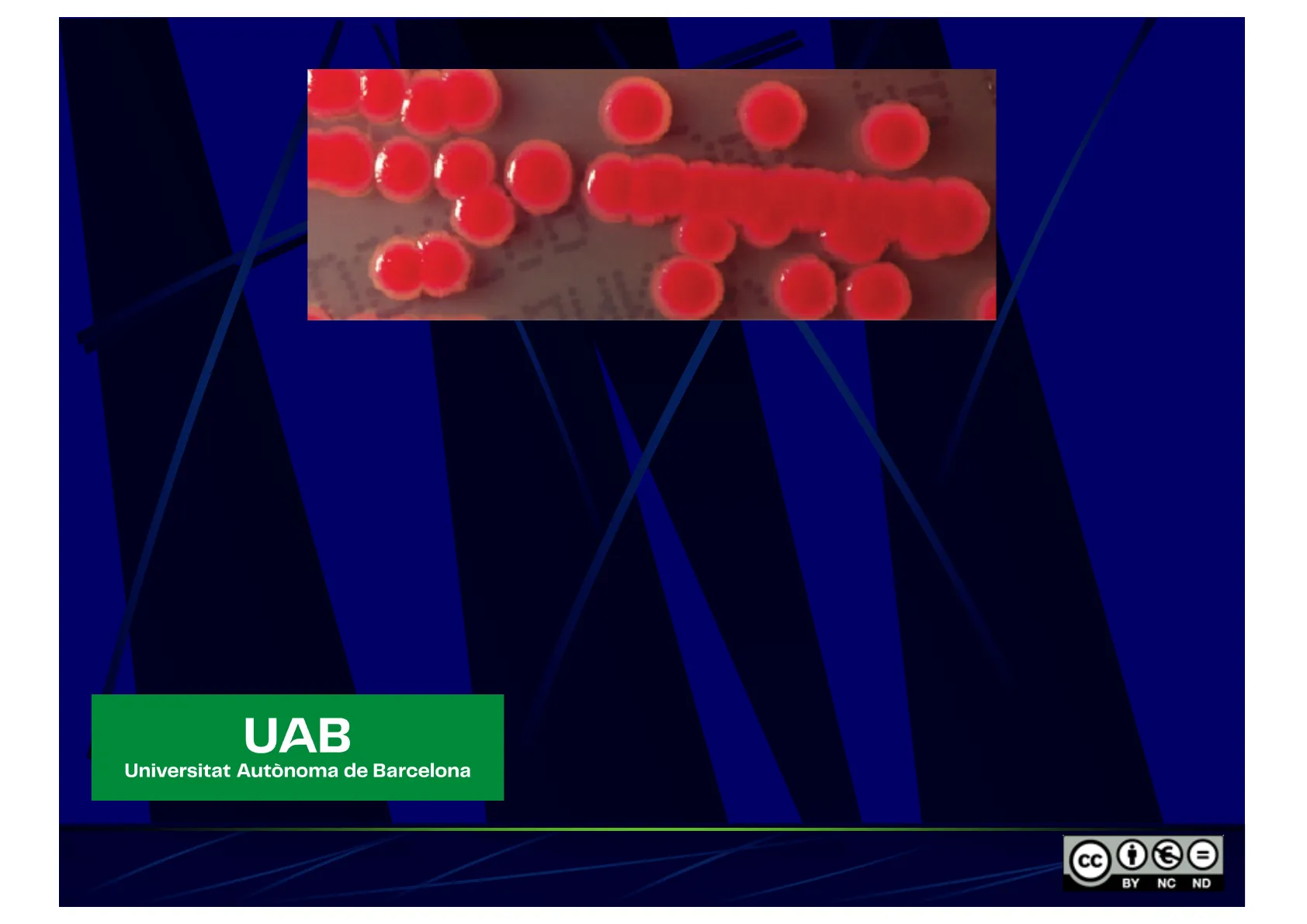
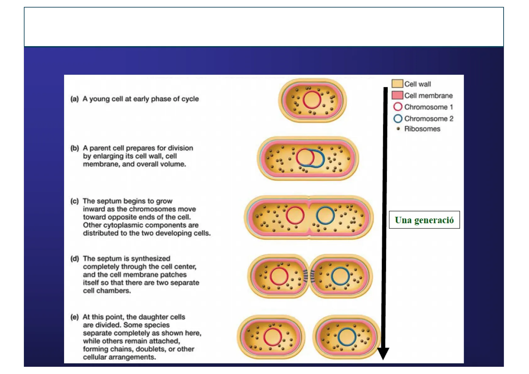
Divisió binària
(a) A young cell at early phase of cycle (b) A parent cell prepares for division by enlarging its cell wall, cell membrane, and overall volume. (c) The septum begins to grow inward as the chromosomes move toward opposite ends of the cell. Other cytoplasmic components are distributed to the two developing cells. (d) The septum is synthesized completely through the cell center, and the cell membrane patches itself so that there are two separate cell chambers. (e) At this point, the daughter cells are divided. Some species separate completely as shown here, while others remain attached, forming chains, doublets, or other cellular arrangements. Cell wall Cell membrane Chromosome 1 Chromosome 2 . Ribosomes Una generació
El cicle cel·lular dels procariotes
Replicació i repartiment del cromosoma
OriC Origin of replication Bacterium Cells divide Replication begins Chromosome Terminator Replisome 20 Chromosomes separate Origins separate Cell elongates as replication continues
El cicle cel·lular dels procariotes
Replicació i repartiment del cromosoma MreB
MreB (homologa a l' actina) forma filaments en espiral que participen en la determinació de la forma cellular i poden servir per a la partició dels cromosomes MinCD i d' altres proteïnes oscilen al llarg de la cel·lula impedint que l' anell Z es formi en llocs diferents al centre de la cèl·lula FtsZ: (semblant a la tubulina) es localitza al centre de la cèl·lula i forma l'anell Z
El cicle cel·lular dels procariotes
Replicació i repartiment del cromosoma FtsZ
Minutes Cell wall 0 - 0 Cytoplasmic membrane Nucleoid MinE + 20 > 40 Divisome complex 60 FtsZ ring 1 Septum - Nucleoid 80 MinE
El cicle cel·lular dels procariotes
Models de divisió cel·lular
- Equal products of cell division: Binary fission: most bacteria
- Unequal products of cell division:
- Simple budding: Pirellula, Blastobacter
- Budding from hyphae: Hyphomicrobium, Rhodomicrobium, Pedomicrobium
- Cell division of stalked organism: Caulobacter
- Polar growth without differentiation of cell size: Rhodopseudomonas, Nitrobacter, Methylosinus
El cicle cel·lular dels procariotes
Cicle cel·lular de Hyphomicrobium
Swarmer cell with subpolar to lateral flagellum (one to three) Young bud New nucleoid moving into hypha Hypha forming 1um Hypha lengthens more and produces another bud.
El cicle cel·lular dels procariotes
Cicle vital de Caulobacter
1pm Pili synthesis Swarmer cell Flagellar rotation Flagellum shedding Completion of cytokinesis Stalked cell Stalk formation
El cicle cel·lular dels procariotes
Bacteris amb micelis
Chain of spores Agar surface (a) 10 M (b) Streptomyces coelicolor 10 sim
El cicle cellular dels procariotes
Bacteris amb micelis M. T. Madigan
M. T. Madigan (a) David A. Hopwood (b)
El cicle cel·lular dels procariotes
Mixobacteris
Myxospores Wall of sporangiole Sporangiole Myxospores Slime envelope Myxospores Stalk Sporangiole Fruiting body of Myxococcus Fruiting body of Polyangium Fruiting body of Stigmatella (a) (b) Myxococcus fulvus (c) Myxococcus stipitatus (d) Chondromyces crocatus
El cicle cellular dels procariotes
Fruiting-body i formació de mixospores
Fruiting-body and myxospore formation Mound of cells Fruiting body Hans Reichenbach Myxospores Swarming and aggregation Slime trails Adventurous motility Chemical induction Germination Hans Reichenbach Vegetative cycle Outgrowth of vegetative cells Figure 15.42 Life cycle of Myxococcus. Dormant myxospores germinate under favorable conditions to yield vegetative cells. Vegetative cells exhibit adventurous motility characterized by gliding (lower inset, Myxococcus fulvus cells, 0.8 pm diameter). When nutrients are limiting, cells aggregate into swarming colonies (upper inset, Myxococcus xanthus colony on agar, 10 mm diameter) exhibiting social motility mediated by type IV pili and twitching. Cells eventually aggregate to form fruiting bodies, within which some vegetative cells develop into myxospores. I Social motility
El cicle cel·lular dels procariotes
Mixobacteris Myxococcus xanthus
oh 8 h 24h 12h Cicle de vida de Myxococcus xanthus
¿Non has encontrado lo que buscabas?
Explora otros temas en la Algor library o crea directamente tus materiales con la IA.
