Cells Structure and Function: Prokaryotic and Eukaryotic Cells
Slides about Cells Structure and Function. The Pdf, a presentation for high school Biology, explores the fundamental differences between prokaryotic and eukaryotic cells, detailing their structure, function, and division mechanisms.
See more34 Pages


Unlock the full PDF for free
Sign up to get full access to the document and start transforming it with AI.
Preview
Structure and Function
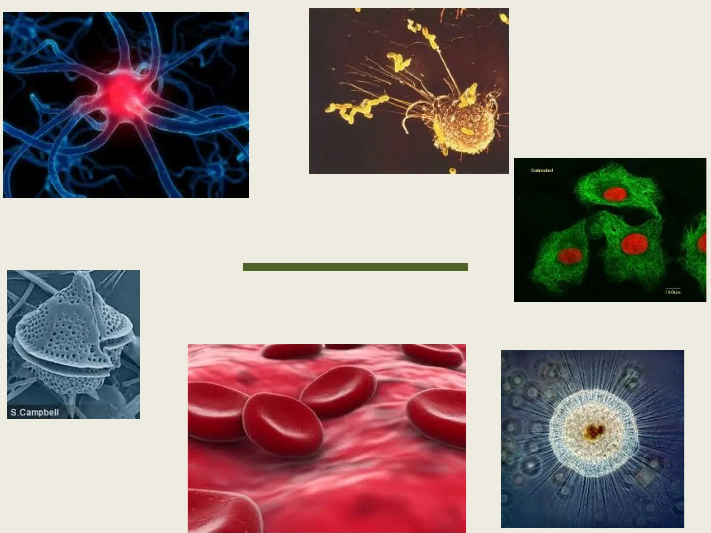
S.CampbellWhy aren't humans made of one giant cell instead of trillions of cells?Why there are not giant cells
- Cells need to pass materials, like nutrients and wastes, into and out of the cell.
- The "doorway" is the cell membrane.
- In order for a cell to survive, there must be enough cell membrane to pass sufficient nutrients into the cell and enough waste materials out of the cell.
- When cells reach a certain "critical size", their surface area (cell membrane) to volume (cell contents) ratio is too small to allow enough molecules to pass in and out of the cell.
- At this point, the cell will divide by mitosis.
3Prokaryotic v Eukaryotic Cells - Amoeba Sisters
Types of Cells
Prokaryotic Cells
- Have one circular molecule of DNA that floats freely in the cytoplasm
- No nucleus
- No organelles except for ribosomes (and some have a flagellum)
- Have a cell wall but it differs in composition from a plant cell wall.
- Divide by binary fission or conjugation.
- Molecules move into and out of the cells by diffusion. Ex. Bacteria, Archaebacteria
Eukaryotic Cells
- DNA is joined with histones to form chromosomes.
- Chromosomes lie in a membrane-bound nucleus.
- Cells contain many different membrane-bound organelles.
- Cells divide by mitosis and meiosis. Molecules move by diffusion, facilitated and active transport. Ex. Plants, Animals, Fungi, Protists
5Types of Cells - Nucleus Medical Media (7:21)
Cell Membrane
- Controls what enters and leaves the cell.
- Acts as a barrier to separate the cell contents from the extracellular fluid.
- Composed of a phospholipid bilayer embedded with proteins (fluid mosaic model).
- Consistency of light oil. Carbohydrate group of glycoprotein Carbohydrate group of glycolipid Extracellular surface of membrane Membrane splits into layers in freeze-fracture electron microscopy. Cholesterol Proteins Intracellular surface of membrane Cholesterol molecules insert themselves into the lipid layer. Lipid tails form the interior layer of the membrane. Phospholipid heads face the aqueous intracellular and extracellular compartments. Copyright @ 2007 Pearson Education, Inc., publishing as Benjamin Cummings. Fig. 3-4
Cytoplasm
- Consistency of "gel".
- Composed mostly of water.
- Also has protein strands, fats, sugars, mineral and other metabolic compounds suspended within.
- Cytoplasm may move in either a circular pattern (cyclosis) or in a linear pattern (cytoplasmic streaming).
- Contains the cytoskeleton of the cell.
8Cyclosis and Cytoplasmic Streaming in a plant cell
Nucleus
- Control centre of the cell, regulates all cell functions.
- Stores the genetic information that is passed on to the next generation.
- Surrounded by TWO nuclear membranes, which have nuclear pores. Nuclear pores Nuclear envelope Chromatin (condensed) Nucleus Nucleolus O O O C O C O Cisternae
10Nuclear envelope (Outer nuclear membrane Nuclear pore Inner nuclear membrane Rough endoplasmic reticulum Chromatin Nucleolus Ribosomes
- Contains one or more nucleoli. Each nucleolus produces ribosomal RNA, which joins with proteins to form the ribosomes.
- The ribosomal subunits exit the nucleus through the nuclear pores, which are about 100nm in diameter. These pores also allow proteins to enter the nucleus so that the ribosomal subunits can be formed.
Chromatin and Chromosomes
Chromatin and Condensed Chromosome Structure Telomere Nuclear Pore Solenoid Chromatin Fiber 1 Nucleosomes- -Centromere DNA - Helix Chromatin -Arm 1 Histones Condensed Chromosome Figure 1 Chromatin is the DNA and proteins of a non-dividing cell, which has a threadlike, grainy appearance. Chromosomes are the rod-like structures that the DNA and proteins form during cell division.
Ribosomes
Ribosome Large subunit Small subunit Ribosome
- Ribosomes are manufactured by the nucleoli.
- They consist of two subunits of RNA and proteins.
- Ribosomes may float freely in the cytoplasm OR they attach to the endoplasmic reticulum.
- Protein synthesis occurs at the ribosomes.
Endoplasmic Reticulum
Three-Dimensional Endoplasmic Reticulum Nuclear envelope Nucleus Ribosomes Rough endoplasmic reticulum Smooth endoplasmic reticulum
- A system of membranous channels and flattened vesicles (saccules) that connects to the nuclear membrane.
- Called Rough ER if there are attached ribosomes.
- Called Smooth ER if there are no ribosomes.
Rough Endoplasmic Reticulum (RER)
- Rough ER looks "dotted" under the microscope. These "dots" are ribosomes.
- Proteins synthesized at ribosomes in the RER are exported from the cell.
- The proteins made here have a sugar chain added to them in the ER, forming glycoproteins.
- Proteins leave the RER in vesicles and move to the Golgi Apparatus Vesicle budding from Ribosome Vesicle rough ER
Smooth Endoplasmic Reticulum (SER)
Mitochondrion Space inside smoth ER
- Smooth ER has NO attached ribosomes.
- Smooth ER synthesizes phospholipids, which are used to form more cell membrane.
- There is lots of SER in the testes, and it produces testosterone.
- SER in the liver produces peroxisomes.
- SER in the Adrenal Cortex gland produces various sex hormones.
Peroxisomes
- Membrane-bound vesicles formed in the SER of the liver.
- Contain hydrolytic enzymes which can break down fats and other harmful substances.
- These enzymes are formed from "free-floating" ribosomes and then transported into the peroxisome by carrier proteins.
- The enzymes in the peroxisome break down molecules into Hydrogen Peroxide (H,O2), which is then immediately broken down into water (H20) and oxygen (O2) by the enzyme Catalase. Anatomy of the Peroxisome Actin Microfilament Network Stress Fibers Nucleus Peroxisomes Animal Cell Micrograph Figure 2 Plasma Membrane Lipid Bilayer Figure 1 Urate Oxidase Crystalline Core
Golgi Apparatus
50 KG
- Found in the cytoplasm.
- Looks like a "stack of pancakes"; really 3 - 20 saccules.
- Receives proteins from the RER and lipids from the SER, both of which arrive in transport vesicles. These molecules move from the inner face toward the outer face of the Golgi. 5 Vesicle from ER "Receiving" side of Golgi apparatus New vesicle forming "Shipping" side of Golgi apparatus Plasma membrane
18Golgi Apparatus
- The Golgi "packages and processes" molecules. For instance, glycoproteins have their sugar chains modified.
- Molecules are put into secretory vesicles to be removed from the cell by exocytosis.
- The Golgi also makes Lysosomes, membrane-bound vacuoles which contain hydrolytic enzymes. The job of the lysosome is to break down macromolecules which are brought into a cell by endocytosis.
Endomembrane System
Smooth ER Nuclear envelope Transport vesicle from ER Nucleus Lysosomes Rough ER Golgi complex Vesicle Plasma membrane
- Proteins made at the RER are packaged into Transport Vesicles to be carried to the Golgi Apparatus.
- The Golgi modifies the molecules which are then packaged into Secretory Vesicles to be carried to the cell membrane.
- The protein is removed from the cell by exocytosis.
Vacuoles and Vesicles
- Larger membrane-bound sacs
- Contain food, waste or pigment molecules or enzymes (if they are lysosomes)
- Form a large central vacuole in plant cells which is full of water, as well as sugars and salts
- Smaller membrane bound sacs
- Can be used to transport molecules around the cell (transport and secretory vesicles)
Mitochondria
- Organelle which is responsible for producing energy for the cell in the form of molecules of ATP.
- Energy is produced when glucose is broken down during the process of Cellular Respiration. CH 0 + 602 6CO + 6H O + energy 2 2 glucose + oxygen -> carbon dioxide + water + ATP
22Inner Membrane Outer Membrane Cristae Matrix
- Consists of a highly folded inner membrane, a central matrix, and an outer membrane.
- The folds of the inner membrane are called cristae, and these folds house the respiratory enzymes of cellular respiration. more folds more SA- more enzymes more ATP energy!
Chloroplast
- Organelle in plant cells which is responsible for synthesizing sugar molecules to use as food.
- The sugar glucose is produced in the process of Photosynthesis in the presence of sunlight. 6CO + 6HO 2 2 CHO + 60 2 carbon dioxide + water glucose + oxygen
24· Consists of an outer membrane which encloses inner membranes called Thylakoids and an inside space called the Stroma. Chloroplast Granum Outer Membrane Lumen Inner Membrane Stroma Thylakoids The thylakoids are stacked into units called Grana (one is granum), and the grana are connected by membranes called Lamellae.
Cytoskeleton
The cytoskeleton is a network of filaments and tubules that run throughout the cell cytoplasm, from the nucleus to the cell membrane. Plasma Membrane Endoplasmic Reticulum Microtubule Mitochondrion Ribosomes Microfilaments and Intermediate Filaments
26five functions:
- Maintain cell shape.
- Assist with cell movement (amoeboid movement)
- Movement of cell organelles within the cell.
- Anchors the cell organelles and cell membrane.
- Assists with the separation of the chromosomes along the spindle apparatus during cell division.
27What makes up the cytoskeleton?
Microtubules
Tubulin dimer a-Tubulin ß-Tubulin MICROTUBULES Cross section Microtubule
- Hollow tubes ~25nm in diameter
- Formed from 13 rows of the globular protein tubulin
- Form the spindle apparatus to which the chromosomes anchor during cell division
- Help maintain the shape of the cell and act as tracks along which cell organelles can move in the cell
Actin Filaments (Microfilaments)
ACTIN FILAMENTS (MICROFILAMENTS) 0 pm
- Two chains of globular actin monomers, twisted into a helix and forming long, thin, flexible fibres, 7nm in diameter
- Occur in bundles or networks, acting in a structural role beneath the cell membrane
- Involved in amoeboid movement (pseudopod formation) and muscle contraction in animal cells.
- Works with the protein MYOSIN in muscle cells to produce movement.
Intermediate Filaments
INTERMEDIATE FILAMENTS
- 8-11nm in diameter
- Made of fibrous polypeptides (like keratin in the skin)
- Found associated with nuclear and cell membranes
- Able to assemble and disassemble Fibrous polypeptide 10nm Buzzle.com Animal Cell Micrograph Nucleus Keratin Intermediate Filament Network Figure 2
31
Can’t find what you’re looking for?
Explore more topics in the Algor library or create your own materials with AI.
