Introduction to Histology: Cell Structure and Tissue Types
Slides about Introduction to Histology, covering cell structure and the four basic tissue types. The Pdf, a university-level Biology presentation, details cellular organelles and their organization, with an example focusing on the digestive system.
See more36 Pages

Unlock the full PDF for free
Sign up to get full access to the document and start transforming it with AI.
Preview
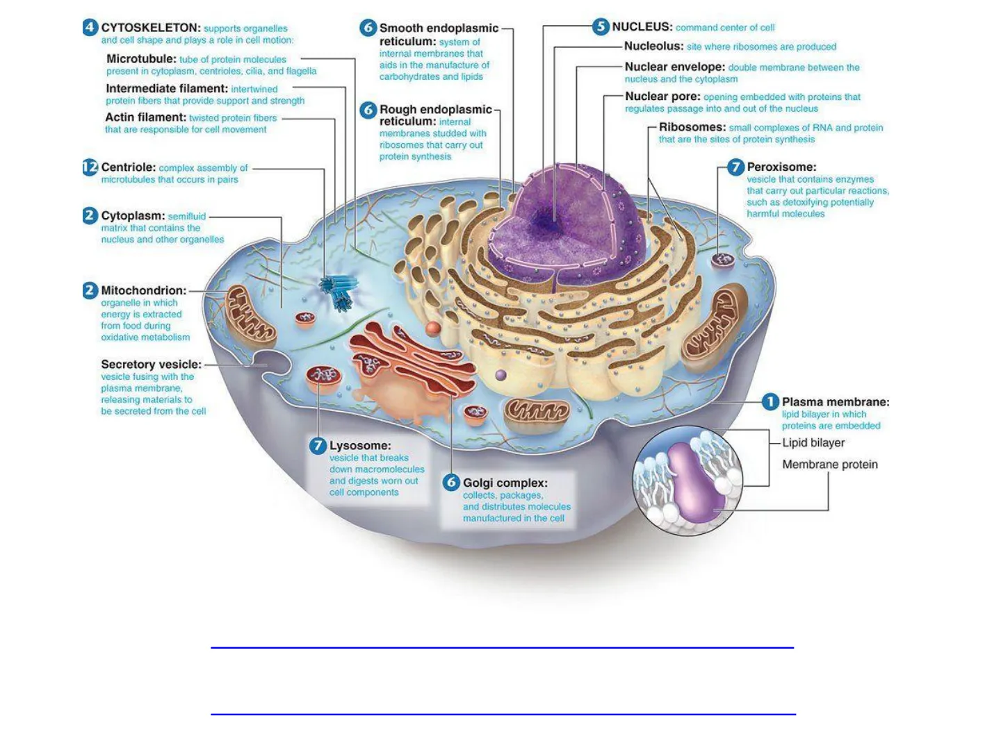
CYTOSKELETON: supports organelles and cell shape and plays a role in cell motion
- Microtubule: tube of protein molecules present in cytoplasm, centrioles, cilia, and flagella
- Intermediate filament: intertwined protein fibers that provide support and strength
- Actin filament: twisted protein fibers that are responsible for cell movement
6 Smooth endoplasmic reticulum: system of internal membranes that aids in the manufacture of carbohydrates and lipids
NUCLEUS: command center of cell
Nucleolus: site where ribosomes are produced Nuclear envelope: double membrane between the nucleus and the cytoplasm Nuclear pore: opening embedded with proteins that regulates passage into and out of the nucleus Ribosomes: small complexes of RNA and protein that are the sites of protein synthesis
12 Centriole: complex assembly of microtubules that occurs in pairs
7 Peroxisome: vesicle that contains enzymes that carry out particular reactions, such as detoxifying potentially harmful molecules
2 Cytoplasm: semifluid matrix that contains the nucleus and other organelles
2 Mitochondrion: organelle in which energy is extracted from food during oxidative metabolism
Secretory vesicle: vesicle fusing with the plasma membrane, releasing materials to be secreted from the cell
1 Plasma membrane: lipid bilayer in which proteins are embedded
7 Lysosome: vesicle that breaks down macromolecules and digests worn out cell components
Lipid bilayer Membrane protein
6 Golgi complex: collects, packages, and distributes molecules manufactured in the cell
https://www.youtube.com/watch?v=URUJD5NEXC8 https://www.youtube.com/watch?v=RKmaq7jPnYM
6 Rough endoplasmic reticulum: internal membranes studded with ribosomes that carry out protein synthesis
FOUR BASIC TYPES OF TISSUE
Tissue : a group of cells that are of similar structure and work together for a particular function.
Tissue Functions
Protection Epithelial tissue: Covers surfaces, lines cavities, forms glands
Movement Muscle tissue: Causes mouvements
Support Connective tissue: Provides support to other tissues, insulates, stores energy reserves
Reception and transmission of signals Nervous tissue: Detects changes, sends and receives impulses
Organ system
A set of organs that interacts to carry out a major body function. The digestive system coordinates the activities of organs, including the mouth, esophagus, stomach, small and large intestines, liver, pancreas, rectum, and anus, to convert ingested nutrients into absorbable molecules and ions, eliminate undigested matter, and help regulate water content of the body.
Organ
Body structure that integrates different tissues and carries out a specific function which, for the stomach, is processing food.
3 2 Stomach Digestive System Stomach Pharynx Liver Colon Mouth and Tongue Esophagus Small Intestine Cecum Gall Bladder Pyforus Pancrean
Tissue Types and Functions
Epithelial tissue: Protection, transport, secretion, and absorption of nutrients released by digestion of food
Connective tissue: Structural support
Muscle tissue: Movement
Nervous tissue: Communication, coordination, and control
1
EPITHELIAL TISSUE
Epithelial Tissue Function
- it covers the external surfaces
- It lines the internal surfaces. Other functions are absorption and secretion.
Wall Mucosa Epithelium Lamina propria Muscularis mucosae Submucosa Submucosal gland Blood vessel Lumen Submucosal nerve plexus Muscularis Inner circular layer Myenteric nerve plexus Outer longitudinal layer Serosa
HOLLOW ORGAN (intestine)
Basement membrane lextracellular matrix)
Free surface of epithelium
Underlying tissue
Cell nuclei
A Simple squamous epithelium (lining the air sacs of the lung)
D Stratified squamous epithelium (lining the esophagus)
B Simple cuboidal epithelium (forming a tube in the kidney)
Layers of dead cells
Rapidly dividing epithelial cells
E Stratified squamous epithelium (human skin)
C Simple columnar epithelium (lining the intestinel
Skin Layers
Epidermis: Stratum corneum Hair Stratum lucidum Stratum granulosum Stratum spinosum Stratum basale Hair shaft Arrector pili Sebaceous gland Sweat duct Sweat gland Subcutaneous fatty tissue Capillary Hair follicle Artery Hair bulb Vein Protects from injury and drying out Dermis
EPITHELIAL TISSUE
Epithelium Basement membrane Connective tissue Capillaries
COMMON FEATURES of Epithelial Tissue
- The epithelium rests on a basement membrane, which separates the epithelial cells and the underlying connective tissue.
- Avascular tissue = it lacks a vascular supply. Cells receive nutrients, water and oxygen through diffusion
- The epithelial cells fit together tightly, with little or no intercellular substance.
- Epithelial cells are polarized. They have a free, or apical, surface, a basal surface and lateral surfaces. The free surface is exposed to the outside or to the lumen; the basal surface rests on the basement membrane
- Epithelial cells regenerate easily
CLASSIFICATION OF EPITHELIAL TISSUE
First name: Number of layers
One layer of cells = Simple Where absorption or diffusion occur
More than 1 layer = Stratified Protection against abrasion, friction and bacteria
Second name: Shape of the cells
= Squamous =Cuboidal = Columnar
Simple Squamous Simple cuboidal Simple columnar
C O 00000 C 0000
Stratified squamous Stratified Cuboidal Stratified columnar
STRATIFIED EPITHELIA
STRATA: Keratinised stratified squamous
squamous spinosum germinativum mitotic figure lamina propria squames
STRATA: corneum granulosum
EPITHELIUM: Keratinised stratified squamous spinosum germinativum
TISSUE / ORGAN: skin (epidermis) mitotic figure dermis
EPITHELIUM: Non keratinised stratified squamous
TISSUE / ORGAN: oesophagus
General features of stratified squamous epithelia
nonkeratinized keratinized Flattened cells in the superficial layer Appear in areas where mechanical insults are frequent Fastness: stratification projections of the connective tissue (papillae) firm cytoskeleton (keratin filaments) firm intercellular junctions (connections) - desmosomes hemidesmosomes at the border of epithelium and connective tissue Avascular and nourished by diffusion of nutrients from capillaries in the connective tissue
Pseudo stratified columnar
Pseudo = False (epithelium looks stratified but it's actually not) Only one layer of cell is present because all of the cells touch the basement membrane
Transitional Epithelium
Can stretch without breaking! umbrella cells
- O Empty bladder = Full bladder = Stratified cuboidal Stratified squamous
Pseudostratified ciliated columnar
0 Simple cuboidal Transitional epithelium Stratified squamous Simple squamous Simple columnar
Membrane Specializations of Epithelia
®
SUMMARY of Epithelial Cells
- Surface epithelia form continuous sheets comprising one or more layers of cells
- Epithelial cells are closely bound to one another
- Epithelial cells are polarized
Epithelial Cells have Polarity
Cilia Apical (free or exposed surface)
Microvilli Apical surface Golgi apparatus Lateral (neighboring epithelial cells) Basal lamina Nucleus Basal (surface anchored to connective tissue) Mocnondria Basolateral surfaces
124
SPECIALIZATIONS OF EPITHELIAL CELLS
Definition: Specialisations of cell membrane are morphologic adaptation for specific functions of particular plasma membrane domains.
Cilia- propel Narrow extracellular space
Microvilli-absorb Apical region of an epithelial cell
Apical specializations
membrane projections of the free surface
Cell junctions Tight junction Epithelium Adhesive belt Desmosome Gap junction Basal region Basal lamina Nerve ending Reticular fibers Basement membrane Connective tissue Capillary
Lateral specializations
JUNCTIONS occur at the interface between two adjacent cells
LATERAL SPECIALIZATIONS
Apical surface
Junction Types and Functions
function seals neighboring cells together in an epithelial sheet to prevent leakage of molecules between them
1 Tight junction
8 Zonula adhaerens joins an actin bundle in one cell to a similar bundle in a neighboring cell
2 Desmosome joins the intermediate filaments in one cell to those in a neighbor allows the passage of small water-soluble ions and molecules
3 Gap junction
Intermediate filaments = Green Actin filaments = Red
Junctional complex
- Tight junction
- Zonula adherens
- Desmosomes
Junctional complex
8 8
Gap junctions
- Plaque-like structure composed of connexons
- A connexon is composed of six subunits of a protein called connexin arranged radially to form a central channel .Exact alignment produces a junction where ions and small molecules can pass.
Plasma membranes Cell 1 Cell 2 The connexons of two cells come together to form a narrow (-1.5 nm) channel through which ions and smal signal molecules can pass. Connexion "Gap" between cells (~2 nm)
Cilia Microvilli Apical surface C C O C Golgi apparatus Nucleus € Mitochondria € Basement membrane Basolateral surfaces
1 - Structure of microvilli
- Small finger-like projection of the apical surface. .Core of cytoplasm with actin filaments. . Actin filaments are anchored to a network of filaments known as "terminal web". At the periphery of the cell the terminal web is anchored to the zonula adherens. .In light microscopy they form the brush border . They are covered by a glycocalix NON-motile; serve to increase surface area
Actin filaments of microvilli Actin filaments of Z. adherens Thickened region of plasma membranes Terminal web Adhesion belt
CILIA
Longer than microvilli. 7-10 um in length
2 - Structure of cilia and flagellum
- Core is called axoneme. 9+2 microtubules organized from a basal body
- Cilia move. Movement is based on the longitudinal sliding of microtubules in the peripheral doublets, powered by ATP.
- They undergo regular synchronous movement: Rapid forward movement (effective stroke) followed by a slower return (recovery stroke)
Ultrastructure of Cilia and Flagella
Axoneme - Outer Doublet Microtubules Central Microtubules Plasma Membrane- -Dynein Arms 1 Basal Body Figure 1
Forward (power) stroke 1 4 5 2 1 6 thm of cilia in are flexible and t the base and muve wuwar'd the tip. Backward (recovery) stroke 10 9 11 8 12 7
Can’t find what you’re looking for?
Explore more topics in the Algor library or create your own materials with AI.
