Human Anatomy Unit 1: The Nervous System and Cranial Nerves
Slides about Human Anatomy Unit 1. The Pdf, a presentation for University Biology students, details the general organization of the human nervous system, including the brain, spinal cord, and cranial nerves, with key neuroanatomy definitions.
See more42 Pages

Unlock the full PDF for free
Sign up to get full access to the document and start transforming it with AI.
Preview
General Organization of the Nervous System
ANATOMIA HUMANA
Unit 1
1
1. The general organization of the nervous system and anatomical terminology
Central Nervous System (CNS)
- Brain: Located in the skull
- Spinal cord: encircled by the vertebral column bones
Peripheral Nervous System (PNS)
All the nervous tissue outside the central nervous system (CNS). This nervous tissue connects
the CNS with the peripheral structures of the body. It includes nerves (cranial and spinal),
ganglia, plexuses and sensory receptors.
Brain and Spinal Cord Anatomy
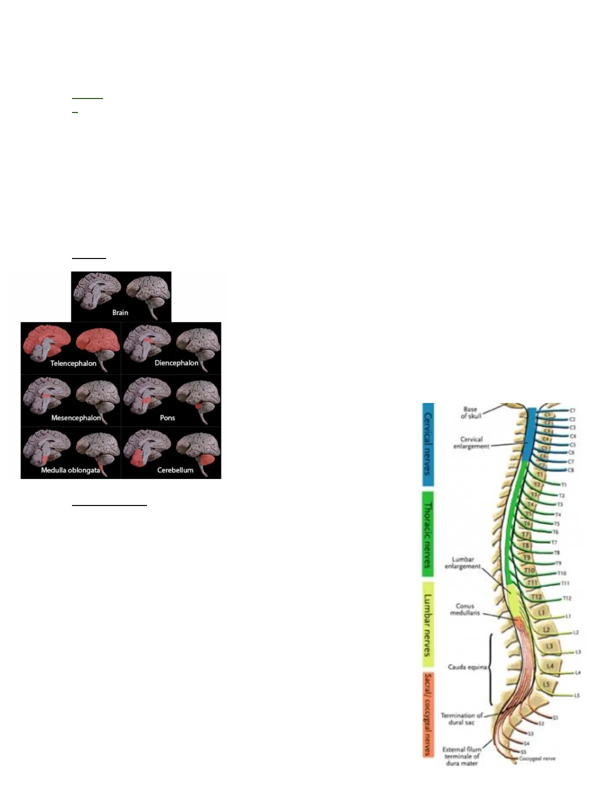
Brain Structures
BRAIN
Brain
Telencephalon
Diencephalon
Mesencephalon
Pons
Medulla oblongata
Cerebellum
Spinal Cord Overview
SPINAL CORD
The spinal cord starts rostrally in the foramen magnum as an
extension of the medulla oblongata.
It is a segmented structure with:
- 8 cervical segments
- 12 thoracic segments
- 5 lumbar segments
- 5 sacral segments
- 1 coccygeal segments
The spinal cord ends caudally with the cauda equina (horse tail) and
the filum terminale. Spinal nerves serving the upper or lower limbs
coalesce to form the brachial or lumbar plexuses respectively.
A plexus, therefore, refers to a network of peripheral nerves.
The fibers of these plexuses are redistributed into the
peripheral nerves.
Brain Regions
TELENCEPHALON the biggest part of the brain outer part
DIENCEPHALON middle and below it is the brainstem
- The Mesencephalon
- Pons
- Medulla oblongata
CEREBELLUM is surrounded by pons.
Base
of skull
Cervical
enlargement
CA
F
12
"
13
T2
Thoracic nerves
17
18
19
Ty
T10
-110
111
T12
T12
Comus
medullaris
L2
Cauda equina
LA
15/
Termination of
dural sac
51
"2
55
External filum
terminale of
dura mater
Coerygral nerve
Cervical nerves
Lumbar
enlargement
Lumbar nerves
Sacral/ coccygeal nerves- The Foramen Magnum has a hole that permits the Medulla Oblongata to connect with
the spinal cord.
- Conus medullaris is the end (poi) form there it comes out the (horse stail) cauda equina
and the filum terminale - Plexus: nerves that are concentrated in an area
Neuroanatomy Vocabulary and Cell Types
Gray and White Matter
NEUROANATOMY VOCABULARY
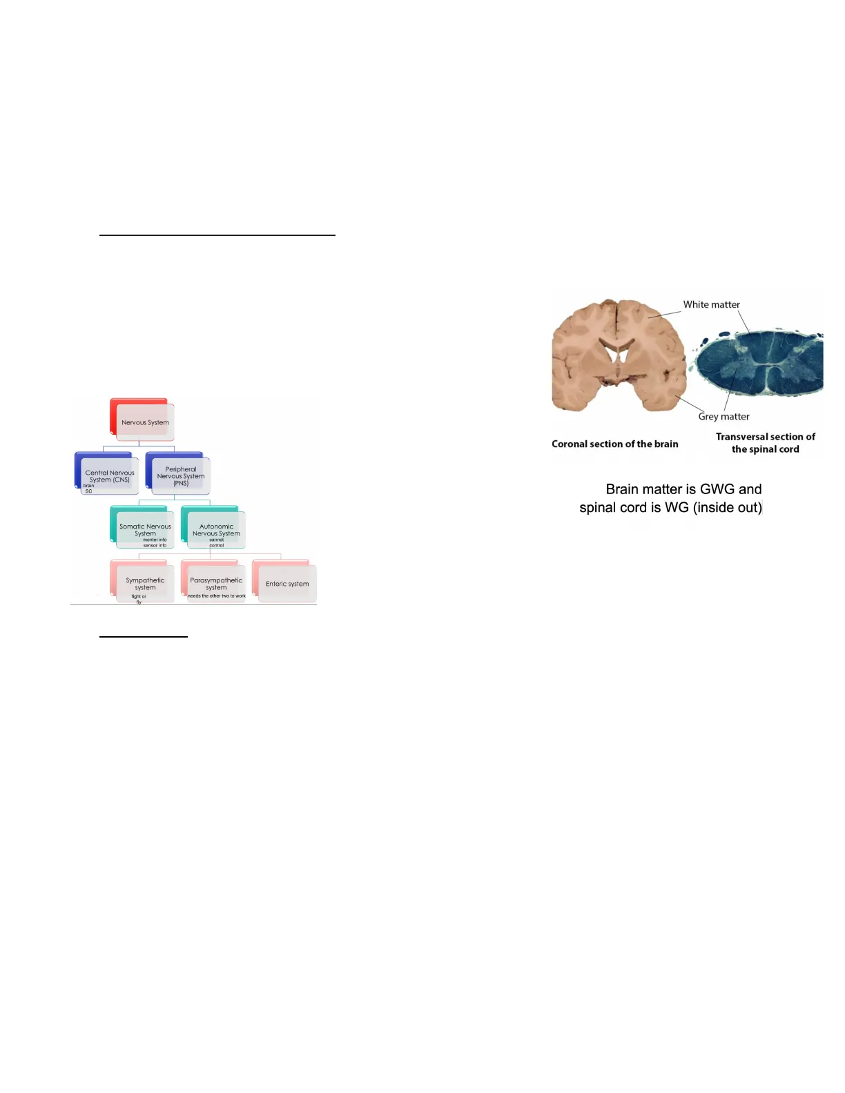
Gray matter: CNS regions rich mainly in neuron cell bodies but also dendrites, unmyelinated
axons and glial cells. For example, the central region of the spinal cord and the cerebral cortex.
(Axon. Myelin sheath is the production of the axon)
White matter: is the pale part of the CNS.
Its color is due to the presence of axon myelinated fibers.
(dendrites and soma)
Nervous System
Central Nervous
System (CNS)
brain
SC
Peripheral
Nervous System
(PNS)
Somatic Nervous
System
monter info
sensor info
Autonomic
Nervous System
cannot
control
Sympathetic
system
fight or
Parasympathetic
system
needs the other two to work
Enteric system
White matter
Grey matter
Coronal section of the brain
Transversal section of
the spinal cord
Brain matter is GWG and
spinal cord is WG (inside out)|
Neuron Types
NEURONS 2
- Unipolar: Has a small axon and doesn't have dendrites coming from the
soma. Ej: Photoreceptive neurons (can detect light) - Bipolar: Has a unique dendrite and axon. Ej .: Photoreceptive neurons of
the eye. - Pseudounipolar: A cell that has a peripheral and central projection. The
soma sits beside the axon. They connect structures. Ej .: Sensitive
afferences. - Multipolar: A big nucleus, loads of dendrites and a long axon. Ej .: In the
nervous system
A neuron is the individual anatomical, physiological, genetic, and trophic unit of the nervous
system.- Oligodendrocyte (create the myelin sheath only in the central nervous system they surround
the axon.)(oligodendrocytes are inside the glial cells) vs. Schwann cells (peripheral nervous
system)
- Nodes of Ranvier: the space between myelin sheath
Neuroimaging and Anatomical Planes
Planes and Terminology
PLANES, AXES ND NEUROIMAGING 3
The neuroanatomical terminology aims at defining, placing and orienting each part of an
organism.
Coronal
Sagittal
Horizontal
The localization or position of any anatomical structure is referred to three orthogonal planes:
horizontal (also called transverse), sagittal (also called medium) and coronal (also called
frontal).
Magnetic Resonance Imaging (MRI)
- Principle: Uses magnetic fields and radio waves to create detailed images of brain
structure. - Advantages: High spatial resolution, non-invasive, no radiation.
- Applications: Diagnosis of tumors, structural abnormalities.
- Image: A detailed black-and-white image of the brain showing internal structures.
Functional Magnetic Resonance Imaging (fMRI)
- Principle: Measures changes in blood flow related to neuronal activity.
- Advantages: Good balance between temporal and spatial resolution, non- invasive.
- Applications: Studying brain activity during cognitive tasks
- Image: Brain activity map with coloured areas indicating levels of activity
Computed Axial Tomography (CT scan)
- Principle: Uses X-rays to create cross-sectional images of the brain.
- Advantages: Fast, effective for visualizing bones and acute injuries.
- Disadvantages: Exposure to radiation.
- Applications: Diagnosis of acute brain injuries, hemorrhages.
- Image: Cross-sectional image of the brain with good contrast between dense (bones)
and soft tissues.
Positron Emission Tomography (PET)
- Principle: Uses radioisotopes to measure metabolic activity of the brain.
- Advantages: Provides precise functional information.
- Disadvantages: Invasive, exposure to radiation.
- Applications: Research on brain metabolism, neurodegenerative diseases.
- Image: Colorful image showing metabolic activity in the brain, with high activity areas
highlighted in warm colors.
CNS Protection: Bones and Meninges
UNIT 2
Bone protection
The CNS is sustained and protected by bones
and membranous meninges. The brain is localized
inside the cranial cavity and the spinal cord in the
vertebral canal of the vertebral column. Inside their
bony coverings, the brain and the spinal cord are
surrounded by three protective membranes, the meninges
The skull supports the structures of the face like nerves,
sensory organs, cavities of the digestive and respiratory
systems and mimic muscles and muscles of mastication.
The skull is composed of two parts: the neurocranium and
the viscerocranium or facial skeleton.
Neurocranium Bones
Neurocranium
The neurocranium is composed of eight bones:
- four are single bones: frontal, occipital, sphenoid and ethmoid bone
- Two are paired bones: parietal and temporal bones.
Frontal Bone Details
Frontal bone
The frontal bone is located in the anterior part of the neurocranium. Two main parts are
distinguished: the superior portion articulates with the two parietal bones by the coronal suture
and the orbital portion that curves downward to make the upper margins of the orbits
Ethmoid Bone Details
Ethmoid bone
The ethmoid bone is located in the anterior part of the base of the cranium, where the cribriform
plate has small apertures through which olfactory nerve fascicles enter the cranium. The crista
galli is a sharp upward projection for the attachment of the falx cerebri.
Frontal bone
Orbital
Frontal bone
portion
4
Sphenoid Bone Details
Sphenoid bone
Bat shape having a centrally placed body with greater and lesser
Wings on its sides. Its located in the middle of the cranial floor
and has a deep depression= the sella turcica which lodges the hypophy:
Coronal suture
oid
Anterior
Posterior
Superior
Crista galli
Cribiform plate
Inferior
Superior Posterior Inferior
view
view
Lateral
view
Frontal
view
Spinal Nerve Origin
Cross Section through Thoracic Vertebra
Meninges
Vertebra of the
Vertebral column
Neurocranium
Viscerocranium or facial
skeleton
AnteriorPartial bones
Parietal Bones Details
The two parietal bones from the sides and roofs of the
cranium and articulate with each other in the midline at
the sagittal structure They articulate with the occipital bone
at the lambdoid structure
Parietal bones
Lambdoid
suture
Parietal bones
Temporal Bones Details
Temporal bones
The temporal bones are at the lateral side of the cranium. Several parts can be distinguished:
the squamous, the petrous and the tympanic parts and the mastoid, the styloid and the
zygomatic processes.
Neurocranium - lateral view
1. Zygomatic process
2. Mastoid process
3. Styloid process
Parietal bone
Frontal bone
Lambdoid
suture
Squamous suture
Temporal bone
Occipital
bori
2
3
Auditory channel
Occipitomastoid
suture
Squamous
part
es
Zygomatic
process
Mastoid
process
Tympanic
part
Styloid
'sidad
Middle cranial fossa
Anterior
cranial
fossa
Posterior cranial fossa
Vertebral Column Structure and Function
Vertebral Column Overview
Vertebral column
The vertebral column is the central bony pillar of the body and has several functions.
- Support for the skull
- Support for the upper limbs
- Support for the thoracic cage
- Transmission of body weight to the lower limbs
- Protection of the spinal cord
The vertebral column laterally has four curves
(kyphosis or lordosis): the cervical, thoracic,
lumbar and sacral curves. When viewed from
the back, the vertebral column runs straight.
7 x cervical vertebrae
12 x thoracic vertebrae
Intervertebral
disc
5 x lumbar vertebrae
5 x sacral
vertebrae
1
4 x coccygeal vertebrae
Cranial Fossae
Neurocranium - cranial fossa
The brain sits over the base of the skull,
which is divided into three cranial fossae.
The anterior cranial fossa is formed by the
frontal, the ethmoid and the sphenoid bones
and contains the two frontal lobes of the cerebrum.
The middle cranial fossa is formed by the tempora
l and the sphenoid bones and hosts the temporal lobes.
The posterior cranial fossa is formed by the occipital
and the petrous parts of the temporal bones. It hosts
the cerebellum and contains the foramen magnum where
the brainstem lodges.
Sagittal suture
Coronal suture
Vertebral Column Composition
Vertebral column composed of 33 vertebrae,
5 sacral fused to form the sacrum and the lower 3
coccygeal fused too. The vertebrae are joint
by fibrocartilage intervertebral discs, making
it a flexible structure
Vertebral column
The two first cervical vertebrae are slightly different,
because they have to sustain the skull but, at the same
time, they have to permit its rotation.C1 vertebra is also
known as atlas and lacks the body.C2 vertebra is also
known as axis, is very robust and has the odontoid
process or dens, that is articulated with the vertebral
arch of atlas allowing the skull rotation.
Dorsal view
Ventral view
Lateral view
Cervical vertebrae
(Cervical curve)
D
0
Intervertebral
discs
Thoracic vertebrae
(Thoracic curve)
Intervertebral
foramina
Lumbar vertebrae
(Lumbar curve)
Sacrum
(Sacral curve)
Coccix
Meninges of the Brain
Meninges Layers
Meninges
Meninges of the brain
The brain, inside the cranium, is surrounded by three protective membranes that are called
meninges.
- The external one is the dura mater and is a hard fibrous membrane.
- The arachnoid mater is the middle layer and is a spider-like membrane composed of
processes called arachnoid trabeculae. - The dura mater and the arachnoid mater are separated by the subdural space.
- The internal meninge is the pia mater, a fine and delicate membrane intimately attached
to the brain. - The arachnoid and the pia mater are separated by the subarachnoid space, which is
filled with cerebrospinal fluid (CSF)
bone
dura mater
arachnoid
arachnoid
trabeculae
- pia mater
glial limiting
membrane
- vessel
- perivascular
space
brain
Bicchiere
Can’t find what you’re looking for?
Explore more topics in the Algor library or create your own materials with AI.
