The Integumentary System: Skin Structure and Superficial Fascia
Slides from University about The Integumentary System. The Pdf, a university-level Biology resource, details the integumentary system, focusing on the skin's structure, including the hypodermis, dermis, and epidermis, and the properties of elastin fibers.
See more56 Pages


Unlock the full PDF for free
Sign up to get full access to the document and start transforming it with AI.
Preview
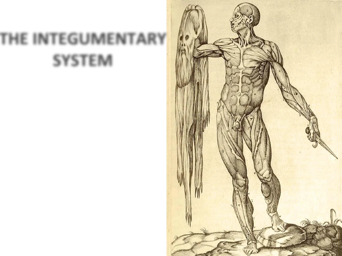
THE INTEGUMENTARY SYSTEM
B M 5 R 5 Dr Valeriano Vinci
WHY DO WE CALL IT "SYSTEM"?
Skin forms the entire external body covering and consists of different tissues, joined to perform specific functions
Facts about Skin
- Weight 12-15% of total body weight
- surface = 1.5-2m2
- 1 cm2 has 70 cm blood vessels, 55 cm nerves, 100 sweat glands, 15 oil glands, 230 sensory receptors
Skin turnover: 25-45 days
AN ORGANIZED GROUP OF TISSUE THAT FORMS A PA
Hair shaft Pore of sweat gland duct Epidermis Arrector pili muscle - Dermis Hair follicle Sebaceous (oil) gland - Hypodermis Hair root Hair follicle receptor Eccrine sweat gland Pacinian corpuscle Adipose tissue Sensory nerve fiber Cutaneous vascular plexus
Sawrosi tuberosa (adenomi sebeon] Clinica Dermatologica - UNINA "Federico II" Dermatology OFRESEARCH www.visualdx.com ( Logical Images, Inc
SKIN FUNCTIONS
1- Protection
- physical barrier that protects underlying tissues from injury, UV light and bacterial invasion.
- mechanical barriers part non specific immunity (skin, tears and saliva).
- prevent water loss.
2- Regulation of body temperature
- hypothalamus reacts to afferent stimuli from the skin
- In case of high temperature or strenuous exercise sweat is evaporated from the skin surface to cool it down.
- vasodilation and vasoconstriction regulate body temperature.
3-Sensation
- nerve endings and receptor cells that detect stimuli to temperature, pain and pressure.
4- Excretion
- sweat removes water and small amounts of salt, uric acid and ammonia from the body surface
5- Blood reservoir
- dermis houses an extensive network of blood vessels carrying 8-10% of total blood flow in a resting adult.
6- Synthesis of Vitamin D (cholecalciferol)
- UV rays in sunlight stimulate the production of Vit. D. Enzymes in the kidney and liver modify and convert to final form; calcitriol (most active form of Vit. D.) aids in absorption of calcium from foods and is considered a hormone.
SKIN STRUCTURE
The human skin (integument) is composed of at least two major layers of tissue: the epidermis and dermis.
Layers Of The Skin
Epidermis
Protects underlying tissue from infection, dehydration, chemicals, and mechanical stress.
Dermis
Contains tough connective tissue as well as different types of cells and glands.
Subcutaneous tissue
Made up of fat and connective tissue, this layer plays many important roles in your body.
Superficial Burn Classification
Epidermis Dermis Subcutaneous tissue CHistirwina, Incorporadid
Superficial Partial-Thickness Burn
Epidermis Dermis Subcutaneous tissue
Full-Thickness Burn
Epidermis Dermis Cheattivive, Incorporates Subcutaneous tissue Healthwise, Incorporated
BURNS
FIRST DEGREE BURN
EPIDERMIS
SECOND DEGREE BURN
DERMIS-
THIRD DEGREE BURN
HYPODERMIS 1 06
EPIDERMIS: KERATINIZED STRATIFIED SQUAMOUS EPITHELIUM WITH FOUR DISTINCT CELL TYPES AND FIVE DISTINCT LAYERS
Dead cells flaking off at the skin surface Stratum corneum Stratum lucidum Stratum granulosum Stratum spinosum Stratum basale Dermis
THE DERMIS
Flexible and strong connective tissue -cells: fibroblasts, macrophages (WBC), mast cells (histamine). - fibers: Collagen - provide tensile strength Elastin - provide for restoration of shape after a deformation - oil and sweat glands origin - nerves, blood and lymphatic vessels
THE DERMIS LAYERS
Hair Dermal papillae Capillary loop of papillary plexus Papillary plexus Papillary layer Reticular layer Cutaneous plexus
Papillary layer:
Loose connective tissue with nipple like surface projection called dermal papilla. provides epidermis: -mechanical anchorage -metabolic support -trophic maintenance Dermo-epidermal innction Epithelial cells Hemidesmosome C Keratin intermediate filament Cytoplasmic plaque (BPAG 1, plectin) Transmembrane anchoring proteins (c64 integrin, collagen XVII/BPAG 2) -Lamina lucida Plasma membrane - Basal lamina -Lamina densa Basal lamina, lamina densa (laminins 1 and 5, collagen IV) Collagen fibril of dermis (collagen III) -Reticular lamina Anchoring fibrils (collagen VII)
Reticular layer:
Merges with the deep aspect of papillary layer and is composed of dense irregular connective tissue - bundles of collagen fibers are thicker - elastic fibers are well represented - orientation of fibers is related to local mechanical forces (skin lines) - include appendages , nerves and vessels
Subcapillary neural Opening (poie) of sweat duet Shat of hair Swort duct Subpspillery vascular Friction ridge Dorral papilas Epidermis I Ders Hypodermis Deep culançous vascular plexus Popikuy lıyor of demis Pacin an corpusche of comes Sweat gand Sesat duet Subcutaneous Seveal gand Hairfollicle Aznice pili muscin Sabesnous gland
Hypodermis:
Loose connective tissue of variable thickness also known as Superficial Fascia - mainly composed of adipose tissue (thermal insulation, shock absorber, storing energy) - mediates attachment of dermis to underlying tissues and mobility of the skin - Superficial Musculo-Aponeurotic System (SMAS)
SUPERFICIAL FASCIA
Skin Subdermal "vertical" fat Fascia superficialis Lymphatic vessels Fascia superficialis fused with premuscular fascia Deep "horizontal" lamellar fat Premuscular fascia Muscle
Elastic Fiber Properties
elastic fiber Elastin fibre - branched and wavy - contain protein elastin - low tensile strength and high deformation range (>100% ) but return to original length STRETCH RELAX single elastin molecule cross-link-
Skin External oblique muscle Superficial fascia- fatty layer (Camper's fascia) Internal oblique muscle Superficial fascia- membranous layer (Scarpa's fascia) Transversus abdominis muscle Transversalis fascia Parietal peritoneum Extraperitoneal fascia
GALEA APONEUROSIS AND INTRINSIC FACIAL MUSCLES
Galea aponeurotica Superior auricular muscle Occipitofrontalis, frontal belly Temporo- parietal Orbicularis oculi ior auricular muscle Nasalis rator labii superioris eque nasi rator labii superioris omaticus minor Occipitofrontalis, occipital belly rbicularis oris Posterior auricular muscle omaticus major Risorius Depressor labii inferioris Mentalis Depressor anguli oris Platysma The epicranial aponeurosis (aponeurosis epicranialis, galea aponeurotica) is an aponeurosis (a tough layer of dense fibrous tissue) which covers the upper part of the cranium in humans and various other animals. In humans, it is attached in the interval between its union with the occipitofrontalis muscle, to the external occipital protuberance and highest nuchal lines of the occipital bone; in front, it forms a short and narrow prolongation between its union with the frontalis muscle or frontal part of the occipitofrontalis muscle.
SUPERFICIAL MUSCULO-APONEUROTIC SYSTEM
- Skin
- Subcutaneous
- Musculo-aponeurotic
- Retaining ligaments and spaces
- Periosteum and deep fascia
Superficial muscular aponeurotic system (SMAS) is an area of musculature of the face. This muscular system
SKIN VASCULAR SUPPLY
SKIN CIRCULATION
- Subepidermal + Dermal «- Subdermal - Plexuses + Subcutaneous Fascia Prefascial and subfascial -Muscle Musculocutaneous artery Regional artery Septocutaneous artery
1 - 1 warm - - hot
SKIN NERVE ENDINGS
Exteroceptors (stimulus outside of body)
- Pacinian corpuscles (lamellated) : deep pressure and stretch
- Meissner's corpuscles (tactile) : light touch, vibration and discriminative touch.
- hair root plexus
- free nerve endings(naked): nociceptors (pain) and thermoreceptors (hot - deep and cold- surface)
- Ruffini's corpuscles: deep pressure
FINGERPRINTS
What are fingerprints?
Found on fingers, palms, toes, soles of feet. Composed of ridges (hills) and furrows (valleys) - Depending on Dermal papillae distribution. ▪ ▪ . Friction ridge skin pattern Black = Ridges White = Valleys
FINGERPRINTS DEVELOPMENT
What are fingerprints?
. Develop in early embryonic life. Pattern is based on genetics and on physical compression of the fetus during pregnancy . So identical twins do not have identical fingerprints ▪
FINGERPRINTS PATTERNS
Arch Loop Whorl
1 Opening of sweat gland ducts Epidermal ridge
Cutaneous "spots"
Nevi (sing. Nevus), generally known as moles, are clusters of melanocytes that appear as small, dark spots.
Types of Nevi
Normal Ephelides (freckle) Lentigo Junctional naevus Compound naevus Intradermal naevus Blue naevus
- One melanocyte to six basal cells
- No increase in number but increase in pigment
- Increased numbers
- Nests of naevus cells
- Nests in dermis but cells get smaller with depth
- Naevus cells only in dermis
- Nodules of dendritic cells deep in dermis
The ABCDEs of Detecting Melanoma
A Asymmetry
NORMAL Symmetrical
MELANOMA Asymmetrical
B Border
Borders Are Even
Borders Are Uneven
C Color
One Color
Multiple Colors
D Diameter
Smaller Than 1/4 Inch
Larger Than 1/4 Inch
E Evolving
Ordinary Mole
Changing in Size, Shape and Color
SKIN APPENDAGES
Hairs V Glands Nails
THE PILOSEBACEOUS UNIT
The structure consisting of hair, hair follicle, arrector pili muscle and sebaceous gland.
Tsc SG Epidermis SS SB Basement membrane Sebaceous gland Permanent segment Dermis Fibroblasts Arrector pili muscle Bulge Outer root sheath Cycling segment Matrix Dermal papilla Hypodermis
Functions of Hairs
V Minor role in humans Protection Thermoregulation V Sensory
Characteristics of Hairs
Present over almost the entire body surface num > 600/cm2 lenght <1 >1 mm width 0.005 ->0.6 mm Vellus hair: fine hairs Terminal hair: hair; axillary and pubic region Distribution depends on: · Sex · Race · Hormones (sex , thyroid, suprarenal, pituitary)
Hair Follicle Structure
Follicle the whole structure surrounding hairs. Bulb is the enlargement at the end of the follicle and houses the germinal layer. Papilla (nipple like) is located in the bulb and is where the blood supply nourishes the hair. Upper part: migrated cells layer during differentiation Hair bulb (papilla) Germinal matrix: closely packed pluripotential cells Keratinocytes Melanocytes Langherans cells
Hairs Structure
Medulla
the inner most layer may even be absent (especially in fine or light blonde hair)
Cortex
provides strength, moisture, colour and texture
Cuticle
outermost layer protects the cortex a hard shingle-like layer of overlapping cells, like scales
SHAFT - ROOT - BULB Bulb contains matrix - inductive mesenchymal cells - melanocytes Hair papilla is at the base of the follicle and contains blood vessels and nerves
Hair Cuticle
Overlapping cuticle cells on the surface of hair
Cortex
Long, slender cells packed like a bundle of sticks
Medulla
A slender, hollow tube in the center of large, coarse hairs
Can’t find what you’re looking for?
Explore more topics in the Algor library or create your own materials with AI.
