Endocrine Pancreas and Pancreatic Hormones, Unicamillus Presentation
Slides from Unicamillus International Medical University in Rome about Endocrine Pancreas and Pancreatic Hormones. The Pdf explores the endocrine pancreas and its hormones, with a focus on anatomy and functions, including paracrine and endocrine effects of insulin and glucose transporter proteins, for University Biology students.
See more59 Pages

Unlock the full PDF for free
Sign up to get full access to the document and start transforming it with AI.
Preview
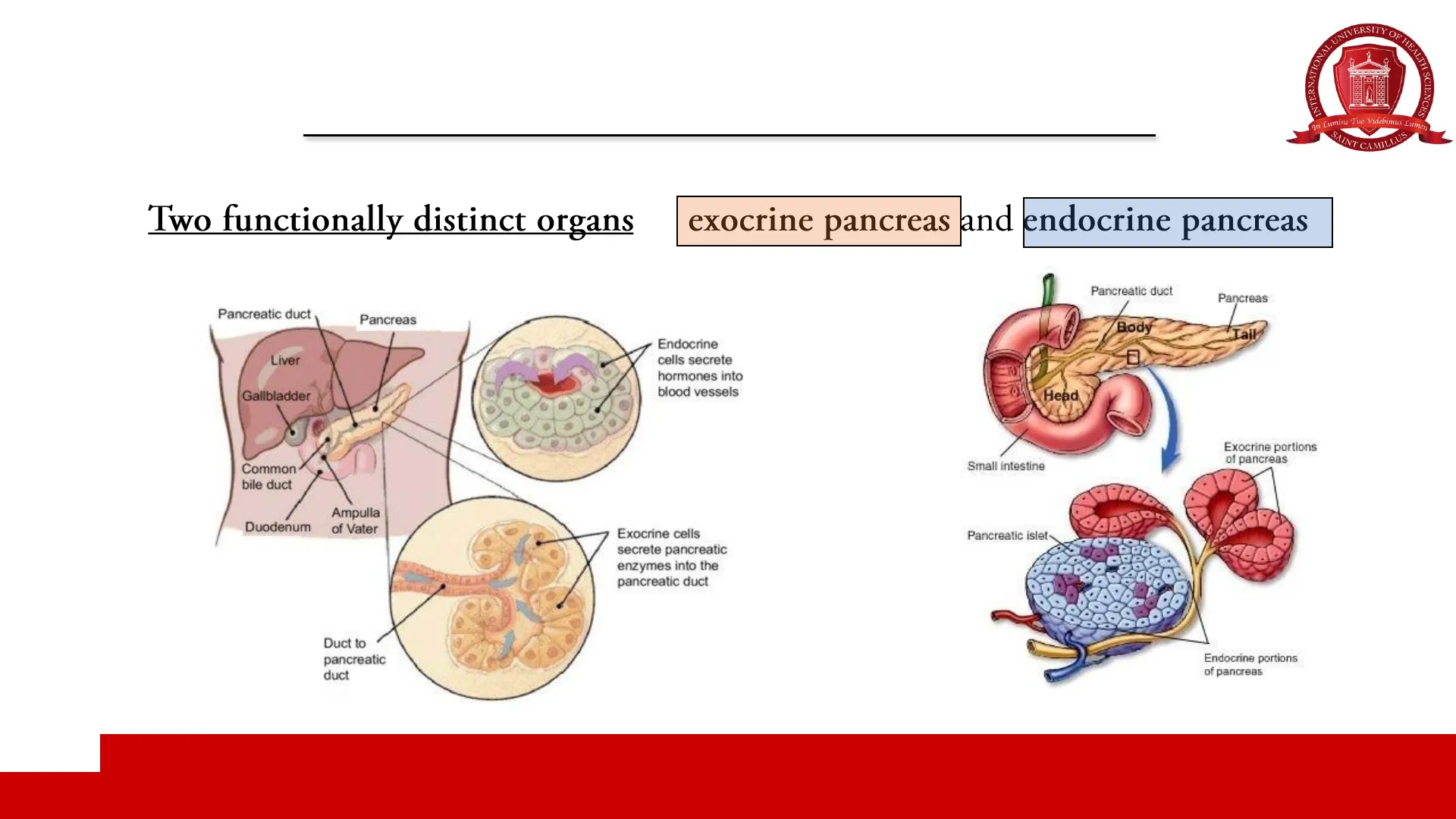
Pancreas Anatomy
OF HE
INTER
In Lumine Tuo Videbimus Lumen
SAINT
IT CAMILLUS
Two functionally distinct organs
→
exocrine pancreas and endocrine pancreas
Pancreatic duct
Pancreas
Liver
Endocrine
cells secrete
hormones into
blood vessels
Gallbladder
Common
bile duct
Duodenum
Ampulla
of Vater
Exocrine cells
secrete pancreatic
enzymes into the
pancreatic duct
Duct to
pancreatic
duct
Mitra 2018 - Journal of Gastrointestinal and Hepatic Surgery
Pancreatic duct
Pancreas
Body
Tail
Head
Exocrine portions
of pancreas
Small intestine
Pancreatic islet
Endocrine portions
of pancreas
TH SCIENCES
Exocrine Pancreas
OF HE
INTER
In Lumine Tuo Videbimus Lumen
SAINT CAMILLUS
a
b
Pancreatic
hormones
Pancreatic islets
TAIL
BODY
Common bile duct
NECK
Digestive
enzymes
HEAD
Duodenal papilla
Duodenum
Pancreatic duct
Acinar cells
The exocrine pancreas (comprised of acinar,
centroacinar and ductal cells) accounts for
approximately 90% of the adult pancreatic mass.
A ORGANIZATION OF THE PANCREAS
Intralobular
ducts
Common
bile duct
Pancreas
Main pancreatic
duct
Intercalated
duct
Interlobular
duct
Acinus
Main
pancreatic
duct
B PANCREATIC ACINAR CELL
Golgi
Intralobular
duct
Pancreatic duct epithelial cell
Centroacinar cell
Pancreatic acinar cell
Mitochondrion
Rough ER
C PANCREATIC DUCT EPITHELIAL CELL
Mitochondria
000
Acinus
Lobule
Zymogen
granules
Intercalated
duct
0
O
TH SCIENCES
Splenic artery
Pancreatic duct
Gallbladder
Lobule
(secretory
unit)
Endocrine Pancreas
Pancreatic Islets of Langerhans
OF HE
INTER
In Lumine Tuo Videbimus Lumen
SAINT CAMILLUS
Splenic artery
Spleen
Pancreas
Pancreatic
hormones:
· Insulin
· Glucagon
Pancreatic islets
Bile duct (from
gall bladder)
Common bile duct
Digestive
enzymes
Pancreatic islet
Alpha cells
Duodenum of
small intestine
Beta cells
Pancreatic duct
Acinar cells
secrete digestive
enzymes
Exocrine acinus
Paul Langerhans
(25 July 1847 - 20 July 1888)
Exocrine pancreas > approximately 90% of the adult pancreatic mass
Endocrine pancreas (about 1 million islets of Langerhans) > 1-2% of the pancreatic mass (weight: about 1 to 2 g in adult humans)
Interstitium (blood vessels, lymphatics, nerves and fibrous connective tissue stroma) > remaining pancreatic mass
TH SCIENCES
Islet Cell Types
Splenic artery
Pancreatic
hormonas
Spleen
Pancreas
Pancreatic islets
. Glucagon
Bile duct (from
gall bladder
Common bile duct
Pancreatic islet
Duodenum of
small irdestina
small intestine
Pancreatic duct
Acinar cols"
societe digestive
enzyTTER
Alpha cells
-Beta cells
Exocrine acinuni
Endocrine pancreas
INTER
IENCES
In Lumine Tuo Videbimus Lumen
SAINT CAMI
ILLUS
Pancreatic islets of Langerhans and islet cell types
Pancreatic islets are richly vascularized and are also innervated by sympathetic, parasympathetic, and sensory neurons.
Human islet of Langerhans
Endocrine cells
TABLE 17-1 Cell types in adult human pancreatic
islets of Langerhans.
Cell Types
Approximate
Percentage of
Islet Volume
Secretory Products
a Cell
25
Glucagon, proglucagon
ß Cell
55
Insulin, C peptide, proinsulin, IAPP,
Ucn3, y-aminobutyric acid (GABA)
8 Cell
10
Somatostatin-14
& Cell
3
Ghrelin
PP cell
5
Pancreatic polypeptide
Insulin
Glucagon
Somatostatin
Cabrera, Ricordi et al. Proc Natl Acad Sci U S A. 2006
Greenspan's, 10th Edition
H SC
Dysfunction and Diabetes Mellitus
Splenic artery
Pancreatic
hormonas
Spleen
Pancreas
Pancreatic islets
. Glucagon
Bile duct (from
gall bladder
Common ble duct
Pancreatie islet
Duodenum of
small intestine
Alpha cells
-Beta cells
Pancreatic duct
Acinar calls
secrete digestive
Exocrine acinuts
Endocrine pancreas
H SC
INTER
IENCES
In Lumine Tuo Videbimus Lumen
SAINT CAMIL
IL LUS
Pancreatic islets of Langerhans and islet cell types
Human islet of Langerhans
Dysfunction of the endocrine pancreas or abnormal responses
to its hormones by target tissues cause serious disturbances in
nutrient homeostasis, including the important clinical syndromes
grouped under the name "diabetes mellitus"
Insulin
Glucagon
Somatostatin
Cabrera, Ricordi et al. Proc Natl Acad Sci U S A. 2006
Glucose Homeostasis
INTER
In Lumine Tuo Videbimus Lumen
IL LUS
Normal fasting plasma glucose values
70-99 mg/dL
Insulin > paramount anti-hyperglycemic
and anabolic hormone
Pancreas
I
Islets of Langerhans
Beta cells
Alpha cells
Insulin
Glucagon
Glucose uptake
Gluconeogenesis
Lipolysis
Skeletal muscle
Liver
WAT
Insulin
I
+
Lipogenesis
Amino acid uptake
Protein synthesis
Ruud J et al. (2017)
H SC
IENCES
SAINT CAMIL
Hormones of the Endocrine Pancreas
H SC
INTER
IENCES
In Lumine Tuo Videbimus Lumen
SAINT CAMIL
IL LUS
Insulin
Amylin
Glucagon
Somatostatin
Ghrelin
Pancreatic polypeptide (PP)
Insulin
Insulin Discovery and Structure
H SC
INTER
IENCES
In Lumine Tuo Videbimus Lumen
SAINT CAMIL
IL LUS
Insulin
H SC
INTE
In Lumine Tuo Videbimus Lumen
SA
NT CAMI
A-chain
1
5
10
15
20
H2N-
Ile
Val
Glu
Gin
Cys
Cys Thr
Ser
Ile
Cys
Ser
Leu
Tyr
Gin Leu
Glu
Asn
Tyr
Cys
Asn -COOH
1
5
10
B-chain
N
HOOC- Thr
Lys
Pro
Thr
Tyr
Phe
Phe
30
25
THE DISCOVERERS OF INSULIN
FREDERICK GRANT
BANTING
1891 -1941
MACLEOD
1876 - 1935
CHARLES HERBERT
BEST
1899 +1970
1892 - 1965
N
Insulin was discovered by Sir Frederick G Banting, Charles H Best and JJR Macleod at
the University of Toronto in 1921 and it was later purified by James B Collip.
In recognition of their life-saving discovery, Banting and Macleod were jointly awarded
the 1923 Nobel Prize in Physiology or Medicine.
> Insulin is a peptide
hormone
consisting of 51 amino acids distributed
among two peptide chains: A-chain (21
amino acids) and B-chain (30 amino
acids).
> Disulfide bonds of cysteine residues
connect the 2 chains.
Insulin > Paramount anti-hyperglycemic
and anabolic hormone
H2N- Phe
Val
Asn
Gin
His
Leu
Cys
Gly
Ser
His
Leu
Val
Glu
Ala
Leu
Tyr
Leu
Val
Cys
Gly
Glu
15
20
Arg
Gly
C
JOHN JAMES
JAMES BERTRAM
COLLIP
IENCES
Insulin Biosynthesis in Beta Cells
H SC
IENCES
An Lumine Tuo Videbimus Lumen
SA
INT CAMI
A
Pdx1
CMofA
(NeuroD1
INS
preproinsulin
(INS) gene
nucleus
preproinsulin
mRNA
rough
endoplasmic
reticulum
C-chain
signal
B-chain
A-chain
preproinsulin
C-chain
B-chain
A-chain
trans-Golgi
network
C-chain
A-chain
B-chain
proinsulin
immature
secretory granules
A-chain
B-chain
insulin
mature
secretory granules
insulin
(hexamer/crystal)
B
glucose
channel
mitochondria
ATPIADP
GK
TCA
several modifying
ion channels
and receptors
shape the
electrial,
endoplasmic
reticulum
oscillatory,
and insulin
exocytic
SST
ACh
glucagon
GLP-1
microtubules
VDCC
ß-cell (insulin)
GABA
a-cell (glucagon)
6-cell (somatostatin)
SNARE protein
complex
released
insulin
Tokarz VL et al. J Cell Biol. 2018
RER
Golgi apparatus
Secretory granules
T1D
X
B chain
A chain
Signal peptidase
PCSK1, PCSK2, CPE
C
Preproinsulin
Proinsulin
Insulin
N-terminal signal peptide
J
C-peptide
Marco Infante et al. Translational Autoimmunity. Volume 4 (Elsevier, 2022)
V insulin
ATP
coupling of B-cells
(and other islet cells)
by gap juctions and
paracrine signals to
promote coordinated,
oscillatory insulin output
Zn2+
><>Ca2+
response
Ca 2 +
cortical
actin
C
To Liver
GLUT
K
The human insulin
gene (INS) is located
on the short arm of
chromosome 11
Proinsulin-Insulin Structure
H SC
INTE
An Lumine Tuo Videbimus Lumen
IL LUS
Structure of human proinsulin, C-peptide and insulin molecules connected at two sites by dipeptide links
PRO
LEU PRO GLN LEU SER GLY ALA GLY
PRO
GLY
GLY
ALA
GLY
LEU
Connecting peptide
GLU
10
GLY
VAL
SER
C chain
GLY
Dipeptide
linkage 31
GLN
VAL
LYS
GLN
ARG
LEU
1
GLY
C chain
(ASP
ILE
-COOH
GLU
VAL
ASN
ALA
GLU
CYS
PHE
GLU 1
GLN
S
TYR
VAL
CYS
ASN
ARG
A chain
GLU
ASN
ARG
THR
B chain
LYS
30
PRO
S
THR
CYS
TYR
GLY
PHE
SER
PHE
HIS
GLY
LEU
ARG
10
GLU ALA LEU TVR LEU VAL CYS GLY GLU
20
C-peptide (31 amino acids)
A chain
(21 amino acids)
Proinsulin
S
B chain
(30 amino acids)
Cleavage
ー ら ー ら ー
+
C-peptide
(No known action)
B chain
SER
ILE CYS SER LEU TYR GLN
S
GLN
S
HIS
1
10
LEU
S
Insulin
Dipep-
tide
linkage
Insulin
(Biologically active)
Greenspan's, 10th Edition
NH2
21
A chain
S-
1
CYS
THR
LEU
B chain
VAL
SA
INT CAMI
IENCES
LEU
GLU
GLN
LEU
C-peptide as a Marker of Endogenous Insulin Secretion
C-peptide: a marker of endogenous
insulin secretion
OF
ALTH
CIENCES
In Lumine Tuo Videbimus Lumen
SAINT CAMILLUS
· Measurement of C-peptide is widely used as a surrogate
marker of endogenous insulin secretory capacity, given
that C-peptide is secreted from pancreatic beta cells at an
equimolar ratio to endogenous insulin, it is not a product of
therapeutically administered exogenous insulin and it is
excreted at a more constant rate over a longer time compared
to insulin.
· Measurement of C-peptide is also useful for the differential
diagnosis of hypoglycemic disorders.
proinsulin
A-chain (21up)
8
4
insulin
C-peptide
(35μμ)
B-chain (30up)
Leighton E, Sainsbury CA, Jones GC. A Practical Review of C-Peptide Testing in Diabetes. Diabetes Ther. 2017 Jun;8(3):475-487
Insulin Properties and Degradation
Insulin
H SC
NTE
An Lumine Tuo Videbimus Lumen
CAM
1
5
10
15
20
H2N- Gly
Ile
Val
Glu
Gin
Cys
Cys
Thr
Ser
Ile
Cys
Ser
Leu
Tyr
Gin Leu
Glu
Asn Tyr
Cys Asn -COOH
H2N- Phe
Val
Asn
Gin
His
Leu
Cys
Gly
Ser
His
Leu
Val
Glu
Ala
Leu
Tyr
Leu
Val
Cys
Gly
Glu
20
Arg
HOOC- Thr
Lys
Pro
Thr
Tyr
Phe
Phe
Gly
N
C
C
30
25
N
Insulin > paramount anti-hyperglycemic
and anabolic hormone
Insulin is a peptide hormone consisting of 51
amino acids distributed among two peptide
chains: A-chain (21 amino acids) and B-chain
(30 amino acids).
Disulfide bonds of cysteine residues connect the 2
chains. An additional intra-chain disulfide bridge
links positions 6 and 11 in the A chain.
Circulatory half-life of insulin: 3-5 minutes
> Insulin is degraded chiefly by insulinases in
liver, kidney, and placenta. A single pass
through the liver removes approximately 50% of
the plasma insulin.
IENCES
1
5
10
15
Physiologic Insulin Secretion
Physiologic Insulin Secretion
24-h profile
H SC
INTE
IENCES
In Lumine Tuo Videbimus Lumen
CAN
Breakfast
Lunch
Dinner
50
Plasma Insulin, mU/L
25
4:00
8:00
12:00
16:00
20:00
24:00
4:00
8:00
Clock Time, h
➢
Basal insulin: small amount of
insulin release during the day in
the fasting state to maintain
normal fasting blood glucose
levels and to inhibit ketogenesis.
It occurs in the absence of
exogenous stimuli (in the fasting
state).
➢
Prandial
insulin: insulin
released from the pancreas to
cover carbohydrates consumed
with meals.
Freeman, 2009
Pancreas
(Pulsatile)
t Insulin
release
3-6 min
Glucose is the most potent stimulant of insulin release
Can’t find what you’re looking for?
Explore more topics in the Algor library or create your own materials with AI.
