Hemoglobin and Myoglobin Structure and Function: Oxygen Transport
Document from Dr. Susan Viselli about Hemoglobin and Myoglobin Structure and Function. The Pdf explores the structure and function of hemoglobin and myoglobin, two fundamental proteins for oxygen transport, including cooperative binding and allosteric effects. This University Biology document was authored by Dr. Susan Viselli.
See more9 Pages


Unlock the full PDF for free
Sign up to get full access to the document and start transforming it with AI.
Preview
Lecture 5
Hemoglobin and Myoglobin: Structure and Function Introduction
Hemoglobin is found exclusively in red blood cells, and its primary function is to transport oxygen. Since oxygen is not very water soluble, and water is the main constituent of blood, an oxygen transport protein must be used. Hemoglobin is composed of four globular protein subunits called globins, and contains a non-protein heme group that reversibly binds oxygen. Each hemoglobin can transport 4 molecules of oxygen (O2). In humans, the average hemoglobin concentration is ~15 g/100 ml. This means there are approximately ~1.5 x 10²⁰ hemoglobin molecules in 100 ml of whole blood. The purpose of hemoglobin is to pick up oxygen at the lungs and deliver it to the tissues. Therefore, hemoglobin must be able to both bind and release oxygen, and at the correct locations. The ability of hemoglobin to reversibly bind oxygen is affected by the partial pressures of O2 (pO2) and of CO2 (pCO2) and the availability of 2, 3-bisphosphogylcerate (2,3-BPG), a product of glycolysis. The interaction of these molecules at one site on the hemoglobin molecule affects the binding of oxygen to other sites of hemoglobin. Collectively these factors (pO2, pCO2, 2,3-BPG) are known as allosteric effectors. Myoglobin is another abundant heme-containing protein. Like hemoglobin, myoglobin binds to oxygen. However, myoglobin is monomeric. It is similar to an individual polypeptide chain of tetrameric hemoglobin. Myoglobin is present in heart and skeletal muscle and is designed to bind oxygen released by hemoglobin at low pO2 of muscle. Myoglobin releases oxygen within muscle cells when the demand for oxygen increases. The binding of oxygen to myoglobin is not influenced by allosteric effectors because it is monomeric and does not have additional oxygen binding sites to be influenced. Understanding the structure and function of myoglobin is useful to gain a better understanding of the more complex hemoglobin and its oxygen binding and delivery capabilities.
Terminal Objective: Hemoglobin Oxygen Binding and Delivery
TERMINAL OBJECTIVE: To understand oxygen binding and delivery capabilities of hemoglobin.
Enabling Objectives
ENABLING OBJECTIVES:
- Describe the overall structure of hemoglobin and compare its T and R forms.
- Explain the mechanisms of cooperative binding of oxygen to hemoglobin and compare the oxygen dissociation curves for myoglobin and hemoglobin when pO2 varies over the range from 0 to 120 mmHg.
- Describe how pO2, pCO2, pH and 2,3-BPG affect the loading and unloading of oxygen to/from hemoglobin and explain why myoglobin is not affected by allosteric effectors.
- Describe the Bohr effect and explain when there is a right or left shift to its equilibrium and how this shift will favor either oxy or deoxy hemoglobin.
- Describe the binding of CO2 and of CO to hemoglobin and explain the recommended treatment of individuals experiencing effects of CO poisoning.
Recommended Reading: Lippincott's Illustrated Reviews: Biochemistry, pages 25-33
I. Hemoglobin Structure and Function
Hemoglobin is found exclusively in red blood cells where it transports oxygen from the lungs to capillaries of the tissues. Anemia is a condition defined by low hemoglobin. Lack of oxygen delivery leads to signs and symptoms including conjunctival pallor and feeling tired. Hemoglobin A (Hb A) is the major form of hemoglobin in adults.
A. Hemoglobin Composition
4 polypeptide chains 2 a and 2 ß chains held together by non-covalent interactions Each chain has:
- regions of a-helical structure
- a hydrophobic heme-binding pocket
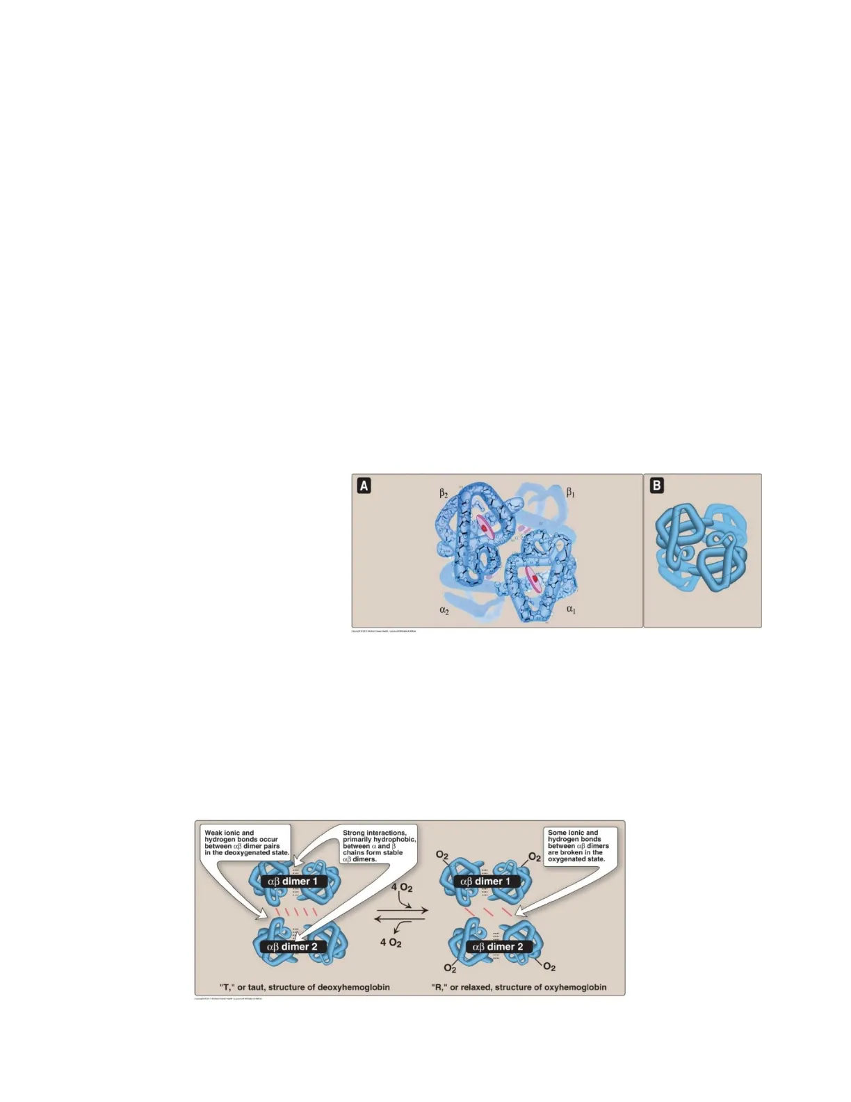
The hemoglobin tetramer is composed of 2 identical dimers: (ap)1 and (ap)2. Chains within each dimer are held tightly together by hydrophobic interactions. The 2 dimers are held together by polar bonds. The weaker polar bonds allow the dimers to move with respect to one another. The 2 dimers occupy different relative positions when O2 is bound to Hb compared to when it is not.
A B2 B
B. Hemoglobin Forms
1. Deoxyhemoblogin - "T" or taut structure The two af dimers interact through ionic and hydrogen bonds that constrain movement of the polypeptide chains. The T conformation is the low-oxygen-affinity form of hemoglobin. 2. Oxyhemoblogin - "R" or relaxed structure The binding of O2 to hemoglobin causes the rupture of some of the polar bonds between the af dimers, allowing movement. The R form is the high-oxygen-affinity form of hemoglobin.
Weak ionic and hydrogen bonds occur between aß dimer pairs in the deoxygenated state. Strong interactions, primarily hydrophobic, between a and B chains form stable aß dimers. Some ionic and hydrogen bonds between aß dimers are broken in the oxygenated state. O2 O2 aß dimer 1 aß dimer 1 - 4 O2 aß dimer 2 aß dimer 2 O2 O2 "T," or taut, structure of deoxyhemoglobin "R," or relaxed, structure of oxyhemoglobin 2 4 02
II. Oxygen Binding
The maximum amount of oxygen that can be carried by blood is determined by the amount of hemoglobin. While there is a small amount of oxygen dissolved in plasma of blood, this amount of oxygen is physiologically insignificant. Hemoglobin can bind 4 O2 molecules, one at each of its 4 heme groups. The degree of saturation of the oxygen-binding sites (known as Y) can vary between 0% when all sites are empty, to 100%, when all sites are full. Pulse oximetry is a noninvasive, indirect method of measuring O2 saturation of arterial blood based on differences in light absorption by oxyhemoglobin and deoxyhemoglobin.
A. Partial Pressure of Oxygen / Oxygen-Dissociation Curve
The primary factor that determines how much oxygen is bound to hemoglobin is the partial pressure of oxygen (pO2 or PO2) in the hemoglobin solution.
- When every oxygen binding site on all the hemoglobin molecules are occupied by oxygen, the blood is considered 100% saturated and the blood cannot carry any more oxygen.
- When half the sites are filled with oxygen, the blood is 50% saturated.
The oxygen-dissociation curve is a plot of Y at different partial pressures of oxygen.
The oxygen dissociation curve for Hb is steepest at the oxygen concentrations that occur in the tissues. This permits oxygen delivery to respond to small changes in pO2. pO2 in tissues pO2 in lungs Myoglobin % Saturation with O2 (Y) 100 Hemoglobin 50 0 OA 40 80 120 Partial pressure of oxygen (pO2) (mm Hg) P50 = 1 P50 = 26 Copyright @ 2011 Wolters Kluwer Health | Lippincott Williams & Wilkins The partial pressure needed to achieve half-saturation of the binding sites (P50) is 26 mm Hg for hemoglobin, but only 1 mm Hg for myoglobin, which contains one heme group and can bind only one molecule of O2.
1. Myoglobin Oxygen-Dissociation Curve
1. For myoglobin (Mb): The oxygen-dissociation curve has a hyperbolic shape, reflecting the fact that myoglobin reversibly binds a single molecule of oxygen. Mb + O2 MbO2 The equilibrium is shifted to the right or left as oxygen is added or removed. Mb is designed to bind oxygen released by hemoglobin at low pO2 in muscle. Mb then releases O2 within the muscle cell in response to demand for O2.
2. Hemoglobin Oxygen-Dissociation Curve
2. For hemoglobin: The oxygen-dissociation curve is sigmoidal, indicating that its 4 subunits cooperate in binding oxygen.
B. Cooperative Binding
Cooperative binding of oxygen by the 4 subunits of hemoglobin means that the binding of an oxygen molecule at one heme group increases the oxygen affinity of the remaining heme groups in the same hemoglobin tetramer. This effect is called heme-heme interaction. Although it is more difficult for the first oxygen molecule to bind to hemoglobin, the binding of additional oxygen molecules occurs with high affinity, demonstrated by the steep upward curve near 20-30 mm Hg. (See figure at bottom of page 3.)
. HE / Hb / Hb O O2. O2 Hb O2 O2 Increasing affinity for O2 D2 O2 Hb O2
III. Allosteric Effects
The ability of hemoglobin to reversibly bind oxygen is affected by the pO2 (heme-heme interactions), by the pH of the environment, by the partial pressure of CO2 (pCO2) and the availability of 2, 3-bisphosphoglycerate (2, 3, -BPG) which are collectively called allosteric effectors. Allosteric means "other site." Allosteric effectors bind to one site on hemoglobin and affect the binding of oxygen to heme groups at other sites on the hemoglobin. (Note - myoglobin is not influenced by allosteric effectors.)
A. Heme-Heme Interactions
The NET effect of specific structural changes initiated at one heme group and transmitted to other heme groups is that the affinity of hemoglobin for the last oxygen bound is ~ 300 times greater than its affinity for the first oxygen bound.
1. Loading and Unloading of Oxygen
The cooperative binding of oxygen allows hemoglobin to deliver more oxygen to the tissues in response to relatively small changes in pO2.
- In the lung, the concentration of oxygen is high and Hb becomes saturated (loaded) with oxygen.
- In peripheral tissues oxyhemoglobin releases (unloads) much of its oxygen for use in oxidative metabolism of the tissues.
LUNGS CO2 is released from hemoglobin. O2 binds to hemoglobin. CO2 O NHỚOO O2 Fe2+ Fe2+ Fe2+ Fe2+ Fe2+ Fe2+ Fe2+ Fe2+ 02 02 NHCOO- Carbaminohemoglobin Oxyhemoglobin CO2 O2 CO2 binds to hemoglobin. O2 is released from hemoglobin. TISSUES Copyright & 2014 Walten Khanes Haam | Lippincem wiliams & Mains 4 02
2. Significance of the Sigmoidal Oxygen-Dissociation Curve
The steep slope of the curve over the range of O2 concentrations between the lungs and tissues permits Hb to carry and deliver oxygen efficiently from sites of high to sites of low pO2. A molecule with a hyperbolic oxygen-dissociation curve, such as myoglobin could not achieve this degree of release within this range of pO2. Instead, it would have maximum affinity for oxygen throughout this pO2 range and would therefore not deliver oxygen to the tissues.
B. Bohr Effect
The release of oxygen from hemoglobin is enhanced when the pH is lowered or when hemoglobin is in the presence of an increased pCO2. This change in oxygen binding is called the Bohr effect.
- Decreased pH and increased pCO2 shift the curve to the right and stabilize the T or deoxyy Hb.
- Increased pH and decreased pCO2 shift the curve to the left and stabilize the R or oxy Hb.
1. Sources of Protons that Lower pH
The concentration of H+ and CO2 in the capillaries of metabolically active tissues is higher than that observed in alveolar capillaries of lungs where CO2 is released into expired air. In tissues, CO2 is converted by carbonic anhydrase to carbonic acid. CO2 + H2O > H2CO3, which spontaneously loses an H+: H2CO3 < > HCO3- + H+ Bicarbonate is THE major blood buffer. The H+ produced by this pair of reactions contributes to the lowering of pH. The differential pH gradient (lungs with higher pH and tissues with lower pH) favors unloading of oxygen in the peripheral tissues and loading of oxygen in the lung.
Decrease in pH results in decreased oxygen affinity of hemoglobin and, therefore, a shift to the right in the oxygen-dissociation curve. pH = 7.6 % Saturation with O2 (Y) 100 pH = 7.2 50 At lower pH, a greater pO2 is required to achieve any given oxygen saturation. 0 0 40 80 120 Partial pressure of oxygen (pO2) (mm Hg) Copyright @ 2014 Wolters Kluwer Health | Lippincott Williams & Wilkins Oxygen affinity of Hb responds to small shifts in pH between the lungs and the oxygen-consuming tissues, making hemoglobin a more efficient transporter of oxygen. 5
Can’t find what you’re looking for?
Explore more topics in the Algor library or create your own materials with AI.
