Introduction to Human Anatomy Course: Concepts and Examination Methods
Document from University about Human Anatomy. The Pdf provides an introduction to the human anatomy course, covering neuroanatomy, splanchnology, microscopic and macroscopic anatomy, and embryology. It is suitable for university-level biology students, offering a clear overview of topics and exam procedures.
See more10 Pages

Unlock the full PDF for free
Sign up to get full access to the document and start transforming it with AI.
Preview
Introduction to Human Anatomy Course
Human Anatomy
Prof. Consalez
26/09/2022
Introduction to the human anatomy course
In the human anatomy course, the first semester has a lot of classes and the fits semester
consists of splanchnology and dynamics of movements meaning. The second semester on
the other hand is neuroanatomy. There are still students who haven't passed
neuroanatomy or have passed it with a very low grade. Neuroanatomy grade accounts for
46% of the final mark. The professor strongly advises us to follow neuroanatomy,
splanchnology and dynamics of movements. He then stresses the importance of attending
the lectures, especially in the second semester.
To encourage students to attend, they decided that students would have to attend each
module by a minimum of 67%. It's no longer like last year when we could reach 67%
attendance in the first semester and be done, this year students have to attend 67% of the
lessons for both the first and second semester. they chose this rule because last year
students followed poorly in the second semester.
Professor Consalez will be our teacher for neuroanatomy and the coordinator of the
anatomy course. He also prefers students to ask questions during the lessons and not be
scared of asking the "wrong" questions. Anatomy is the course with the highest number of
credits (18 in total). The course will be divided into 180 hours of human anatomy lectures
and 40 human anatomy practical including dissection practical at the end of the second
semester that will be very useful for topographic anatomy. The credits of the exams are
coordinated with the European credits transfer system so if anyone wants to move to
another university, they will be acknowledged, even in the United Kingdom.
Aim of the human anatomy course is to provide the morphological, both macro and
microscopic, basis of functions and diseases
Anatomy
of our organism. Human Anatomy course is a
blend of Organogenesis, Microscopic and
Macroscopic anatomy.
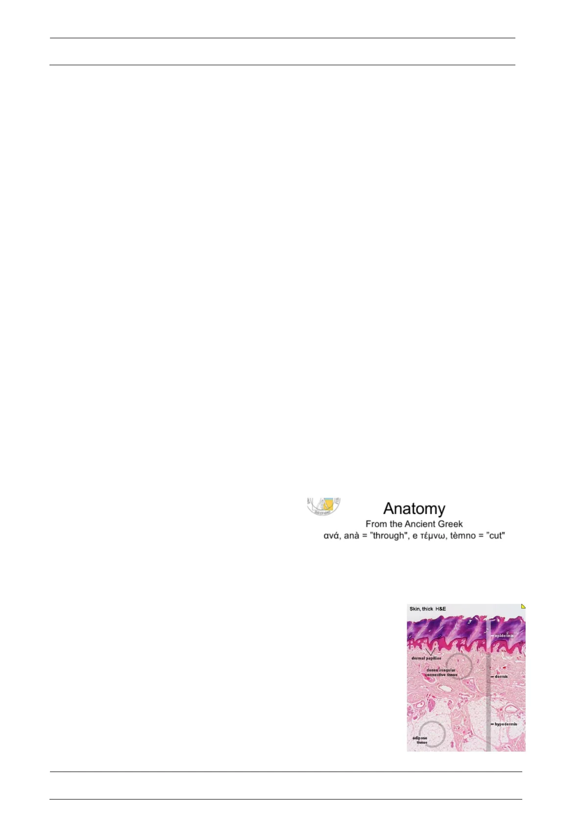
Anatomy Terminology
From the Ancient Greek
ανά, ana = "through", e τέμνω, temno = "cut"
- Organogenesis: How organs and systems of the body
develop. - Microscopic anatomy: How different types of tissues of the
body are assembled to form organs. It also involves the
examination of cells and molecules. (Follow up of
Histology) Learning the features of individual cells that
form a tissue in Histology than understanding how those
tissues make up an organ. (Nevertheless, Microscopic
Anatomy can also examine a cell or tissue as said before.)
It can be said that Microscopic Anatomy divides into
Cytology and Histology.
Skin, thick H&E
epidermis
dermal papillae
dense irregular
connective tissue
- dermis
- hypodermis
adipose
tissue
Author: Onur Can Gormez
Pag. 1/10Human Anatomy
Prof. Consalez
26/09/2022
On the right, you can see an example of microscopic anatomy. How different types of
tissues like the epithelium, the connective tissue, and the adipose tissue assembles to
make up our integuments, meaning our skin, dermis and hypodermis. We are going to
learn the main features of these tissues in histology and we will assemble these
components in microscopic anatomy.
- Macroscopic/Gross Anatomy: Visual knowledge of the body structure. It is the study
of anatomical structures that can be seen by naked eye. Gross Anatomy comprises
of Systemic, Regional, Functional and Applied Anatomy. Macroscopic anatomy can
be subdivided into systematic anatomy where we study an organ system (for
example digestive) from head to toe and topographic anatomy where we take a
given segment of the body and we have to recognize all the structures that make up
that segment. - Systemic Anatomy is the anatomy of the systems inside the body. It provides an
overview of the system throughout the body. - Regional/ Topographic Anatomy is an approach to anatomic study based on
regions, parts and divisions of the body. It emphasises the relationships of systemic
structures such as muscles, nerves and arteries within that area. It may indicate the
relationship of various systemic structures. This is why Systemic Anatomy will be
the later foundation of Topographic Anatomy. - Functional/Physiological Anatomy indicates the functional significance of the
structures, by providing basic structural knowledge provided in Human Anatomy. It
is the study of structure-function relationships. (Studying anatomy in its relation to
function.) In Functional Anatomy, we try to introduce a lot of functional references
when we describe the morphology and give a preview of some diseases, thus we
do not just basically memorise some structures, but we understand the human
diseases and their basis. - Applied Anatomy is also a subdivision of Macroscopic Anatomy, but itself also
comprises of some other subdivisions. Applied Anatomy examines structure-
function relationships in context of related subjects. For example: Radiologic,
Computational, Clinical and Microscopic Anatomy.
Brief Introduction to Embryology
Embryology is the ground work for organogenesis. We will talk about the early
development from fertilisation to gastrulation and the entire embryonic period.
Embryology comes from the
ancient Greek έμβρυον,
embryonic, "the unborn" and -
λογία - logia"-discourse"
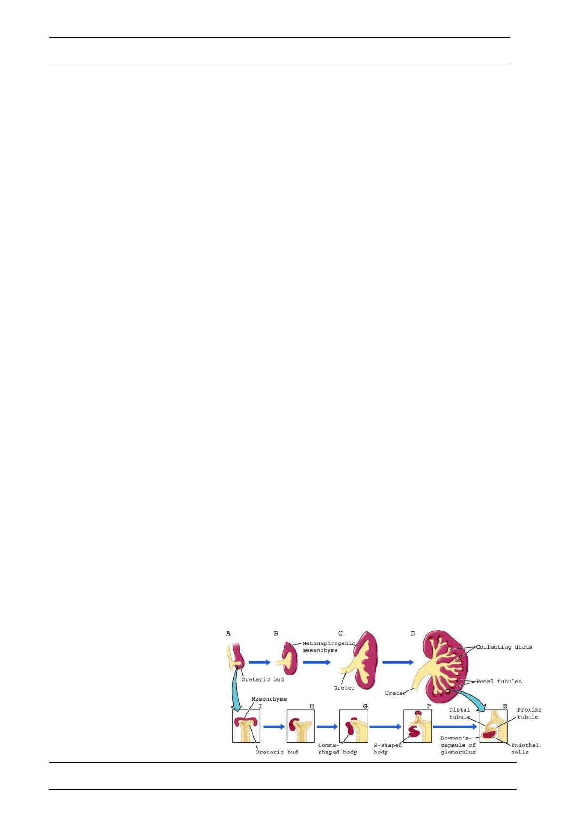
Organogenesis deals with the
development of the urinary
system, as shown here in the
labelled diagram, it also plays
A
B
C
D
-Metanephrogenic
mesenchyme
Collecting ducts
Ureteric bud
Renal tubules
Ureter
Ureter
Mesenchyme
I
H
G
F
E
Distal
tubule
Proxima
tubule
Ureteric bud
Comma-
shaped body
s-shaped
body
Bowman's
capsule of
glomerulus
Endothel:
cells
Author: Onur Can Gormez
Pag. 2/10Human Anatomy
Prof. Consalez
26/09/2022
a role in the cardiovascular, respiratory and digestive systems. Histology provides the
basis to anatomy that provides the basis for physiology.
Neuroanatomy
Neuroanatomy is an unique field of anatomy as there cannot be experimentations on
humans. A lot of information on human neuroanatomy is inferred from animal models such
as rodents and non human primates. It is continuously evolving as there are still a lot of
research on this anatomical discipline.
Neuroradiology imaging will potentially bridge the gap between comparative and clinical
neuroanatomy. (Comparative Anatomy is the comparative study of the body structures of
different species of animals in order to understand the adaptive changes they have
undergone.)
Neuroradiology also gives a lot of important information regarding the function of specific
areas of the brain. (compared with healthy and diseases individuals.)
Human Anatomy Terminology
Human anatomy is the unambiguous description of thousands of (macro- and microscopic)
structures and is impossible without the use of an extensive and highly specialised
terminology. So there is an anatomical terminology used worldwide. (Anatomical
terminology will be covered up by Ottavio Cremona.)
This terminology of Human Anatomy has been standardised worldwide, and there are
databases for anatomical terminology as well as organisations that make sure the
terminology that is used all around the world is standardised.
Anatomy cannot be thought in one shot, and there is a set of information needed in order
to navigate through Human Anatomy. This is why, we are going to learn, repeat the main
information and add more to it. This addition of information to pre-existing one will create
the term "learning through apposition".
We are going to learn anatomy through comparing for example, theoretical vs practical
knowledge , gross vs microscopic anatomy , descriptive vs functional anatomy. Moreover,
problem based learning will also be the foundation of our Anatomical knowledge.
(Associating the disease and problems of the patient with an anatomical basis is referred
as problem based learning.)
Author: Onur Can Gormez
Pag. 3/10Human Anatomy
Prof. Consalez
26/09/2022
Textbooks
Suggested Textbooks
- ANATOMY BAG PLUS (Systemic & Topographic Anatomy & Atlas) pagina 43
- GROSS ANATOMY TEXTBOOK Moore: Clinically Oriented Anatomy
- GROSS ANATOMY TEXTBOOK Gray's Anatomy for Students
Suggested Atlases
- GROSS ANATOMY ATLASes Netter's Atlas (the most used one) 4
- ATLAS of GROSS ANATOMY THIEME's Atlas (has a very good design)
- John Martin Neuroanatomy text and atlas ( Professor suggest this one for
neuroanatomy)
Microscopic Anatomy makes us of some tools such as "The
Aperio System". It is a new way of looking histological slides,
microscopic anatomy. This new system has a very high
resolution for even single cells and components of the tissue,
while these images can be transferred to a monitor. (This gives
the opportunity to look at them wherever you want.) The Aperio
System is a modern and efficient tool for microscopic anatomy.
Webscope
@39496
Here in this picture of a kidney, we have the
maximum resolution where we can see the single
components of the organ. We will be able to use
this tool both in class and at home.
Exams
In the first semester, we will be able to take the dynamics of movements and the
splanchnology written tests. We can take these exams 4-5 times so we will have many
opportunities to pass them. The dynamic of movements and splanchnology exams can be
taken in December while for neuroanatomy the first opportunity will be in July or
September. For human anatomy practicals, there will be a test after the dissection labs
and it is a pass or not exam so no grades only in July. The minimum threshold is 18/30.
The exam will be computer-based with 1 question at a time and no back button. We will
have 60 multiple-choice questions in 72 minutes for each exam.
The final grades are calculated:
Author: Onur Can Gormez
Pag. 4/10Human Anatomy
Prof. Consalez
26/09/2022
0.44 * Splanchnology + 0.12 * Dynamics of movement + 0.44 * Neuroanatomy = 1
Cells of the Central Nervous System
CNS is populated with cells which have soma(cell body) located at one site, but the effect
of neuronal activity is felt sometimes a meter away (even longer in taller people). This is
done by the axons of (motor) neurons.
The metabolic load for this situation is very demanding, but this is how some neurons are
communicating with very distant sites within the body. This property is unique to the
neurons.
Between all cells of the CNS and PNS (peripheral nervous system), 20% of them are
neurons. Some of these neurons project information to very distant locations called motor
neurons, whereas some neurons function locally. These are called "local circuit neurons/
interneurons".
Neurons can have different shapes such as, "bipolar, pseudounipolar and multipolar".
(These will be covered in the next lesson)
Remaining 80% of cells of the nervous system (CNS and PNS) are glial cells. These are
the cells that provide support to the neurons. They are cells that provide axons with its
myelin sheath. There are myelin-forming cells called oligodendrocytes in the central
nervous system and Shawn cells in the peripheral nervous system. There are other
structures in the central nervous system or in the so-called neuron tube such as ventricles,
cells that line these ventricles and produce cerebral spinal fluids. (The professor said that
he will cover this later on).
Haematoxylin & Eosin staining is especially commonly
used by pathologists, in order to understand the tissue
Np
in terms of tumours, neurodegenerations etc. For
example, in this photo you can see the H&E
preparation of the brain. (Haematoxylin is an
G
acidophilic die with blueish properties, whereas Eosin
is a basophilic die with reddish properties.) Neurons
can be distinguished as it has much bigger soma than
other types of cells.(Soma of the neurons can reach 100 microns across in diameters, but
for a typical neuron it is about 20 micrometers in diameters.)
Glial cells such as oligodendrocytes and astrocytes can be distinguished from the H&E
staining. Oligodendrocytes can be recognised by their pale cytoplasm, and they provide
myelin sheets to the axons of the neurons in Central Nervous System.(Schwann Cells-
PNS) Astrocytes are recognised by their nuclei that are compact and dense.(their structure
and function will be covered in the next lesson.)
Author: Onur Can Gormez
Pag. 5/10
Can’t find what you’re looking for?
Explore more topics in the Algor library or create your own materials with AI.
