Special Senses: Hearing, Anatomy of the Ear and Sound Transduction
Slides about Special Senses – Hearing. The Pdf covers the anatomy of the ear, the physiology of hearing, and the process of sound transduction. The Pdf, suitable for university students studying Biology, includes detailed diagrams and step-by-step explanations of how sound waves are converted into electrical signals.
See more12 Pages

Unlock the full PDF for free
Sign up to get full access to the document and start transforming it with AI.
Preview
Structure of the Auditory System
In this lecture we will be covering ........ Structure of the auditory system Physiology of hearing Sound transduction Anatomy of the Ear
GERARD J. TORTORA BRYAN DERRICKSON PRINCIPLES OF ANATOMY & PHYSIOLOGY Organization, Support and Movement, and Control Systems of the Human Body 18TH EDITION VOLUME INTERNATIONAL STUDENT VERSION Chapter 17
- The ear is an engineering marvel because its sensory receptors can transduce sound vibrations with amplitudes as small as the diameter of an atom of gold (0.3 nm) into electrical signals 1000 times faster than photoreceptors can respond to light
- The ear also contains receptors for equilibrium, the sense that helps you maintain your balance and be aware of your orientation in space
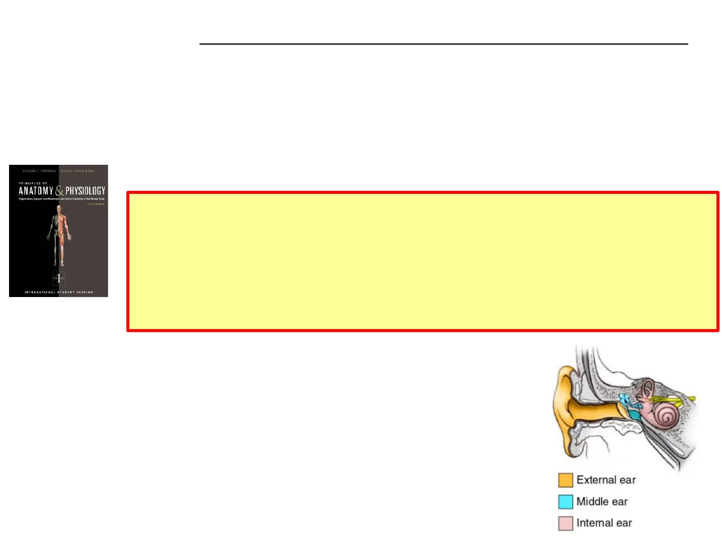
The ear is divided into three main regions:
- external ear - collects sound waves and channels them inward
- middle ear - conveys sound vibrations to the vestibular window
- internal ear - houses the receptors for hearing and equilibrium
External ear Middle ear Internal ear
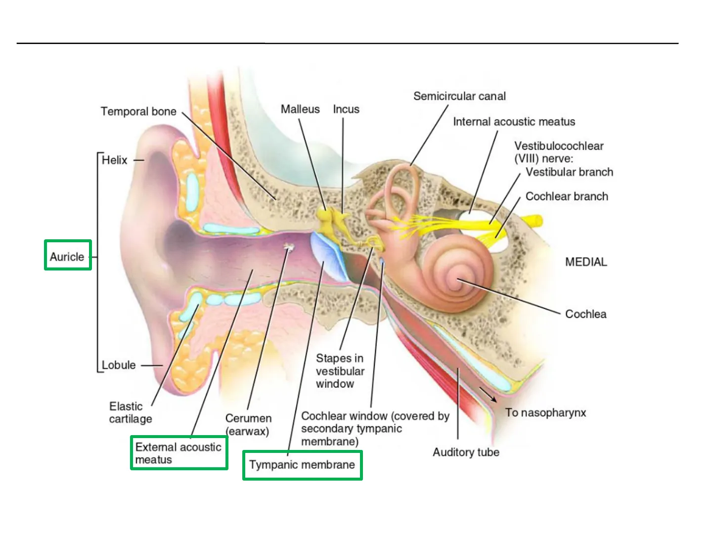
External Ear Anatomy
External ear Semicircular canal Temporal bone Malleus Incus Internal acoustic meatus Helix - Vestibulocochlear (VIII) nerve: Vestibular branch Cochlear branch Auricle MEDIAL Cochlea Lobule Stapes in vestibular window Elastic cartilage To nasopharynx Cerumen (earwax) Cochlear window (covered by secondary tympanic membrane) External acoustic meatus Auditory tube Tympanic membrane
Middle Ear (Tympanic Cavity)
Middle ear (tympanic cavity)
- Separated from internal ear by a thin bony partition that contains two small openings:
- vestibular window
- cochlear window
- Extending across the middle ear are the auditory ossicles;
- Malleus (hammer)
- Incus (anvil)
- Stapes (stirrup)
Superior ligament of malleus Posterior ligament of incus Malleus Incus Stapes in vestibular window Facial (VII) nerve Lateral ligament of malleus Cochlear window Anterior ligament of malleus (cut) Tensor tympani muscle MEDIAL LATERAL Tympanic membrane External acoustic meatus Stapedius muscle Tympanic cavity Auditory tube (a) Coronal section showing location of auditory ossicles in the middle ear
- Anterior wall of the tympanic cavity contains an opening that leads directly into the auditory tube or pharyngotympanic tube, commonly known as the eustachian tube
- Connects tympanic cavity with the nasopharynx - normally closed at its medial (pharyngeal) end - opens during swallowing and yawning to equilibrate air pressure in tympanic cavity
Inner Ear (Labyrinth)
Inner ear (labyrinth) Bony labyrinth (contains perilymph) Membranous labyrinth (contains endolymph) Semicircular canals (contain semicircular ducts): Ampulla of semicircular canal Anterior Utricle Posterior Lateral Vestibule Vestibular window MEDIAL Saccule Cochlea Cochlear duct Ampulla of semicircular duct Stapes in vestibular window Cochlear window Components of the right internal ear
Bony Labyrinth Structure
Bony labyrinth
- series of cavities covering 3 areas
- semicircular canals
- vestibule
- cochlea
- lined with periosteum and contains perilymph which surrounds the .....
Membranous Labyrinth Structure
Membranous labyrinth
- series of epithelial sacs and tubes inside the bony labyrinth
- house the receptors for hearing and equilibrium
- contains endolymph
- high levels of K+
- important for the generation of auditory signals
Cochlea Structure
Cochlea Utricle Stapes in vestibular window Saccule MEDIAL Scala vestibuli Cochlea Scala tympani Cochlear duct Scala vestibuli Vestibular membrane Cochlear duct Basilar membrane K Secondary tympanic membrane in cochlear window Scala tympani Transmission of sound waves from scala vestibuli to scala tympani by way of helicotrema
Cochlea Channels
Divided into three channels:
- Cochlear duct - continuation of the membranous labyrinth (endolymph)
- Scala vestibuli - channel above the cochlear duct and ends at the vestibular window
- Scala tympani - channel below is the scala tympani and ends at the cochlear window
- Both scala part of bony labyrinth (perilymph)
Cochlea Scala vestibuli (contains perilymph) Vestibular membrane MEDIAL Cochlear duct (contains endolymph) Tectorial membrane Spiral organ Spiral ganglion Basilar membrane Cochlear branch of vestibulocochlear (VIII) nerve Scala tympani (contains perilymph) (c) Section through one turn of the cochlea
- Vestibular membrane separates the cochlear duct from the scala vestibuli
- Basilar membrane separates the cochlear duct from the scala tympani
- Resting on the basilar membrane is the spiral organ or organ of Corti
Spiral Organ (Organ of Corti)
Spiral organ - organ of Corti
- A coiled sheet of epithelial cells, including supporting cells and about 16,000 hair cells, (receptors for hearing)
Tectorial membrane Stereocilia Outer hair cell Inner hair cell Supporting cells Cochlear branch of vestibulocochlear (VIII) nerve Basilar membrane Cells lining scala tympani (d) Enlargement of spiral organ
Hair Cells of the Spiral Organ
Inner hair cells (receptors for hearing) - single row - convert the mechanical vibrations of sound into electrical signals Outer hair cells (not hearing receptors) - three rows - increase the sensitivity of the inner hair cells
Spiral organ - organ of Corti Stereocilia at apical tip of each hair cell extend into the endolymph of the cochlear duct
- . Basal ends of inner and outer hair cells synapse with first-order sensory neurons and motor neurons from the cochlear branch of the vestibulocochlear (VIII) nerve
Tectorial membrane Stereocilia Outer hair cell Supporting cells Inner hair cell Cochlear branch of vestibulocochlear (VIII) nerve Basilar membrane Cells lining scala tympani (d) Enlargement of spiral organ
- Ends of the stereocilia of the hair cells are embedded in the tectorial membrane while the bodies of the hair cells rest on the basilar membrane
- Sound waves of various frequencies cause certain regions of the basilar membrane to vibrate more intensely than other regions
Sound Waves Characteristics
Sound waves
- originate from a vibrating object
- alternating high- and low-pressure regions traveling in the same direction
Frequency of a sound vibration is its pitch - entire audible range extends from 20 to 20,000 Hz - most acutely 500 and 5000 hertz (Hz) Intensity (size or amplitude) of the vibration, the loudness - measured in units called decibels (dB)
- An increase of one decibel represents a tenfold increase in sound intensity
- The hearing threshold - the point at which an average young adult can just distinguish sound from silence - is defined as 0 dB at 1000 Hz
15 dB - rustling leaves 30 dB - normal conversation 60 dB - vacuum cleaner 90 dB - nearby motorcycle
Physiology of Hearing
Malleus Incus Stapes vibrating in vestibular window Helicotrema Cochlea Sound waves MEDIAL Perilymph outer hair cell tectorial membrane cells of Hensen -hair bundle cells of Claudius reticular lamina 1 > 2 9 External acoustic meatus 8 Spiral organ Tectorial membrane cells of Boettcher tunnel of Corti basilar membrane cells of Deiters nerve fibres @1997 Encyclopaedia Britannica, Inc. Tympanic membrane Secondary tympanic membrane vibrating in cochlear window Tympanic cavity Auditory tube
- The auricle directs sound waves into the external auditory canal
- Sound waves strike tympanic membrane - vibrates in a frequency dependent manner
- Flows down malleus, incus and stapes
- Movement of stapes vibrates oval window
- Causing changes to fluid pressure in perilymph of scala vestibuli
- Transmitted to scala tympani and then cochlea window
- Pressure waves distort vestibular membrane creating waves in endolymph of cochlea duct
- Causes vibration of basilar membrane moving hair cells of spiral organ against tectorial membrane - bending of stereocilia and generation of nerve impulses
3 4 7 Scala tympani 5 Scala vestibuli 6 Basilar membrane inner hair cell pillar cells Vestibular membrane Cochlear duct (contains endolymph) 8
Sound Transduction Process
Tip link (resting position) Tip link (stretched) Tip link (slack) K* Stereocilium 0 Cation channel (partially open) Hair cell Small amount of K enters the cell Voltage-gated Ca2+ channel Voltage-gated Ca2+ channel Voltage-gated Ca2+ channel Ca2+-o Strong depolarizing receptor potential Ca2+ Ca2+ Hyperpolarizing receptor potential O Synaptic vesicle Neurotransmitter Synaptic vesicle Neurotransmitter First-order auditory neuron First-order auditory neuron (c) Hyperpolarized hair cell
- Inner hair cells transduce mechanical vibrations into electrical signals
- Vibration of basilar membrane causes sterocilium to bend (b) - tip links are stretched and tug on mechano-gated ion cation channel (transduction channel)
- Activation (opening) - allow K+ influx (due to high levels in endolymph)
- Induces depolarisation of hair cell and opening of voltage-gated calcium channels (Ca2+ influx)
- Triggers exocytosis of synaptic vesicles containing neurotransmitter
- Release of neurotransmitter proportional to nerve impulses generated
- If they stretch the opposite direction (c), tip links become slack - closes the transduction channels and the cells hyperpolarise, reducing neurotransmitter release and therefore frequency of action potentials at the auditory nerve fibres
- Cation channel (closed) Hair cell Hair cell Large amount of K* enters the cell K' is not able to enter the cell Weak depolarizing receptor potential Ca2+ Ca2 Synaptic vesicle Neurotransmitter First-order auditory neuron (a) Resting hair cell (weakly depolarized) (b) Strongly depolarized hair cell Stereocilium 000 Stereocilium Cation channel (completely open) K
Auditory Pathway to the Brain
f Primary auditory cortex in temporal lobe of cerebum Medial geniculate nucleus in thalamus Inferior colliculus in midbrain Cochlear branch of vestibulocochlear (VIII) nerve Lateral lemniscus Superior olivary nucleus in pons Cerebellum Cochlear nuclei in medulla oblongata From hair cells of the cochlea, auditory information is conveyed along the cochlear branch of the vestibulocochlear (VIII) nerve and then to the brainstem, thalamus, and cerebral cortex
- Some axons from the cochlear nuclei cross over and ascend the lateral lemniscus tract on the opposite side, and terminate in the inferior colliculus of the midbrain
- Other axons from the cochlear nuclei end in the superior olivary nucleus of the pons
- Differences in the timing of nerve impulses arriving from the two ears at the superior olivary nuclei allow us to locate the source of a sound
- Axons to the primary auditory cortex in the temporal lobe of the cerebrum where conscious awareness of sound occurs
Can’t find what you’re looking for?
Explore more topics in the Algor library or create your own materials with AI.
