Human Anatomy I: Temporal, Parietal, and Ethmoid Bones
Document from Human Anatomy I-prof. Sciamanna G. about Human Anatomy I-Prof. Sciamanna G. The Pdf explores the anatomy of temporal, parietal, and ethmoid bones, detailing their structures, processes, and foramina. This university-level Biology material, produced by Prof. Sciamanna G., includes anatomical illustrations to aid understanding of the cranial bone components.
See more13 Pages

Unlock the full PDF for free
Sign up to get full access to the document and start transforming it with AI.
Preview
Temporal Bones
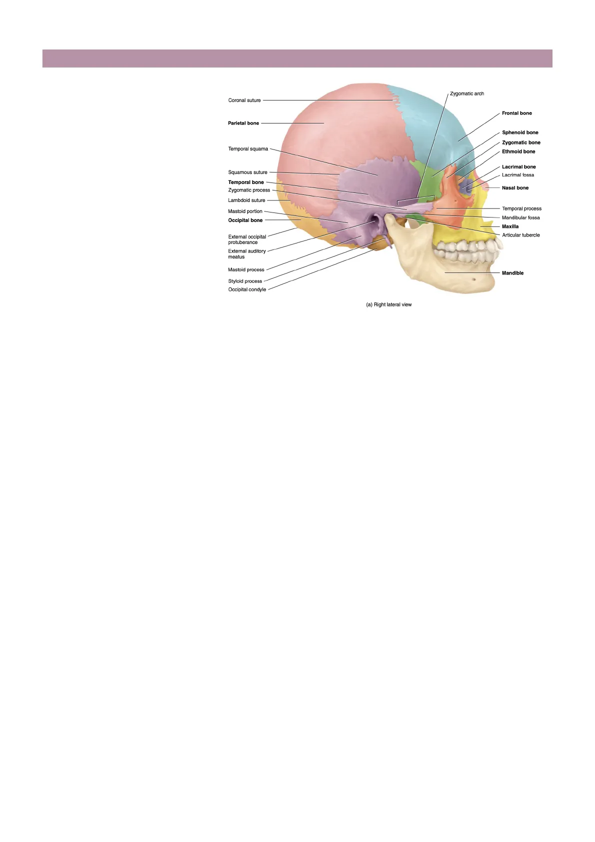
· The temporal squama, the thin, flat portion of the temporal bone that forms the anterior and superior part of the temple (=the region of the cranium around the ear). · Projecting anteriorly from the inferior portion of the temporal squama is the zygomatic process, which articulates (=forms a joint) with the temporal process of the zygomatic (cheek) bone. · On the inferoposterior surface of the zygomatic process of the temporal bone is a socket called the mandibular fossa.
Zygomatic Arch and Related Structures
Zygomatic arch Coronal suture Frontal bone Parietal bone Sphenoid bone Zygomatic bone Temporal squama Ethmoid bone Squamous suture Lacrimal fossa Temporal bone Zygomatic process Lambdoid suture Mastoid portion Mandibular fossa Occipital bone Maxilla External occipital protuberance Articular tubercle External auditory meatus Mastoid process Mandible Styloid process Occipital condyle (a) Right lateral view
The mandibular fossa and articular tubercle articulate with the mandible (lower jawbone) to form the temporo- mandibular joint (TMJ).
Parts of the Temporal Bone
In the temporal bone we can recognize different parts, for example:
- The mastoid portion is located posterior and inferior to the external auditory meatus; there we can find the mastoid process is a rounded projection of the mastoid portion of the temporal bone posterior and inferior to the external auditory meats that serves as a point of attachment for several neck muscles (such as the sternocleidomastoideole).
- The external auditory meatus, the point where we have the connection between the middle and the inner ear; where we have the cochlea and the vestibule-important for some perceptions and balance.
- The internal auditory meatus is the opening through which the facial-controlling facial expressions-(VII) and vestibulocochlear-controlling the receptors for the sound and the balance-(VIII) cranial nerves pass.
- The styloid process projects inferiorly from the inferior surface of the temporal bone and serves as a point of attachment for muscles and ligaments of the tongue and neck.
Associated Structures with Temporal Bone
Other important parts associated with the temporal bone are:
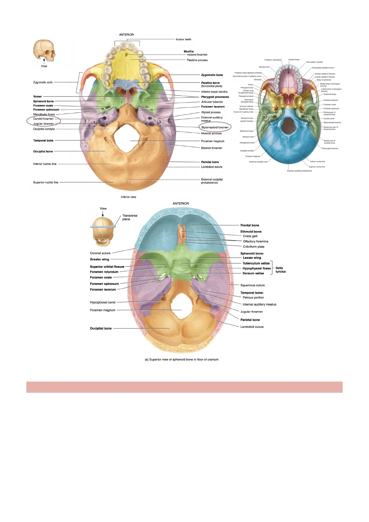
- Between the styloid process and the mastoid process is the stylomastoid foramen, through which the facial (VII) nerve and stylomastoid artery pass.
- Petrous portion At the floor of the cranial cavity is the petrous portion (petrous=rock) of the temporal bone. This portion is pyramidal (having the shape of a pyramid) and located at the base of the skull between the sphenoid and occipital bones; it's the more internal part, forming the floor of the cranial cavity-therefore more visible in the internal part. The petrous portion houses the internal ear and the middle ear, structures involved in hearing and equilibrium. It also contains the carotid foramen, through which the carotid artery passes. Posterior to the carotid foramen and anterior to the occipital bone is the jugular foramen, a passageway for the jugular vein (=one of the most important veins, associated with the venous drainage of the brain), formed by adjacent notches in the temporal and occipital bones.
1 Lacrimal bone Nasal bone Temporal processANTERIOR Incisor teeth Maxilla: Incisive foramen Palatine process Posterior nasal spine Incisive fossa Hard palate (maxilla) View Alveolar arch Hard palate (palatine bone) Posterior nasal aperture (choana), Pyramidal process of palatine bone Hamulus Greater palatine foramen Lesser palatine foramen Body of sphenoid Zygomatic arch Palatine bone Medial plate of pterygold Vome process Pterygoid fossa Greater wing Lateral plate of pterygoid process Inferior nasal concha (of sphenoid bone) Scaphold fossa Vomer Pterygoid processes Sphenoid bone Articular tubercle pterygoid canal Foramen ovale Foramen lacerum Articular tubercle Mandibular fossa Groove for auditory tube Petrous part of Mandibular fossa temporal bone Carotid foramen Styloid process Jugular foramen Carotid canal Jugular foramen Stylomastoid foramen Squamous part of temporal bone Mastoid process Mastoid process Temporal bone Foramen magnum Hypoglossal canal Basilar part of occipital bone Mastoid foramen Occipital bone Occipital condyle Foramen magnum Parietal bone Inferior nuchal line Lambdoid suture Superior nuchal line External occipital protuberance Inferior view ANTERIOR View 1 Transverse plane Frontal bone Ethmoid bone: Crista galli Olfactory foramina Cribriform plate Coronal suture Sphenoid bone: Greater wing Tuberculum sellae Superior orbital fissure Hypophyseal fossa Foramen rotundum Dorsum sellae Foramen ovale Foramen spinosum Squamous suture Foramen lacerum Temporal bone: Petrous portion Hypoglossal canal Internal auditory meatus Foramen magnum Jugular foramen Parietal bone Lambdoid suture Occipital bone (a) Superior view of sphenoid bone in floor of cranium
Parietal Bones
- It has a more regular organization compared to the other bones.
- The two parietal bones are large, quadrilateral (four-sided) bones that form the greater portion of the sides and roof of the cranial cavity.
- Each bone articulates with five other bones; largely interconnected with the rest of the skull.
- The external surface of each parietal bone bears a pair of low ridges, the superior and inferior temporal lines.
- These lines mark the attachment of the temporalis muscle, a large muscle that closes the mouth. The smooth parietal surface superior to these lines is called the parietal eminence.
- The internal surfaces of the parietal bones retain the impressions of cranial veins and arteries that branch inside the cranium.
2 External occipital crest Inferior nuchal line Superior nuchal line External occipital protuberance Pterygoid process Opening of Foramen lacerum Foramen ovale Foramen spinosum -Foramen spinosum Styloid process External auditory meatus Stylomastoid foramen Occipital condyle Mastoid notch Pharyngeal tubercle Lesser wing Sella turcica Zygomatic bone (horizontal plate). The external surface is smooth and convex.
Important Features of Parietal Bones
- Important features: Superior temporal line which forms an arch that travels between the frontal and occipital borders of the parietal bone. The superior temporal line represents the attachment point of the temporal fascia. Superior to this line is the epicranial aponeurosis (=flat tendon connected to one of the muscles covering the skull of the cranium), while inferior to it is the temporal fossa.
Coronal suture Zygomatic arch Temporal process of zygomatic bone Zygomatic process of temporal bone Frontal Bone Supra-orbital foramen Parietal Bone Sphenoid Superior temporal line Inferior temporal line Frontonasal suture Nasal Bone Squamous suture Lacrimal Bone Lambdoid suture Lacrimal groove Occipital Bone Ethmoid Temporal Bone Maxilla External acoustic meatus Infra-orbital foramen Mastoid process Zygomatic Bone Styloid process Mandible Mental foramen Mental protuberance Coronoid process
Inferior temporal line which forms an identical arch to the previous one but located more inferiorly. It represents the origin of the temporal muscle-one of the muscles involved in mastication, moving from the temporal fossa and attaching to the coronoid process of the mandible, controlling the movement of the mandible . Parietal eminence (=small protuberance) which is located centrally on the external surface of the parietal bone. It marks the origin of ossification of the parietal bone.
ANTERIOR View Zygomatic bone Frontal bone Coronal suture Sagittal suture Parietal bones Sutural bone Occipital bone (b) Superior view
Occipital Bone
- Forms the posterior part and most of the base of the cranium. it appears as a platelike bone with a somewhat triangular shape. Its inferior portion is thick.
- It is divided into four parts arranged around a large opening, the foramen magnum: the basilar part, two condylar parts and the squamous part.
3
- The basilar part (1) sits anterior to the foramen magnum and adjacent to the petrous part of the temporal bone.
- The condylar parts are located lateral to the foramen magnum. They comprise two oval- shaped prominences (occipital condyles) that articulate with the first cervical vertebra (atlanto-occipital joint).
- Superior to each occipital condyle on the inferior surface of the skull is the hypoglossal canal -where we find the exit from the skull of the hypoglossal nerve, that has as targets the oral cavity and the tongue (*glossal=something referring to the tongue)
ANTERIOR Incisor teeth Maxilla: Incisive foramen Palatine process Sagittal suture View Zygomatic bone Zygomatic arch Palatine bone (horizontal plate) Inferior nasal concha Vomer Pterygold processes Sphenoid bone Articular tubercle Foramen ovale Foramen lacerum Foramen spinosum Styloid process Mandibular fossa 1 Carotid foramen External auditory meatus Occipital bone Squamous part Supreme nuchal line Petrous part Mastoid foramen Occipital bone Superior nuchal line External occipital protuberance Inferior nuchal line Mastoid foramen Mastoid process Superior nuchal line Occipital condyle Temporal bone Palatine bone Inferior view Mandibular foramen Sphenoid bone, pterygoid process Maxilla, palatine process Incisive foramen Mandible Teet! Coronal suture Frontal bone Sphenoid bone Sella turcica: Sagittal plane Dorsum sellae Tuberculum sellae Incus Hypophyseal fossa View Frontal sinus Ethmoid bone: Crista galli Parietal bone Cribriform plate Temporal squama Squamous suture Lambdoid suture Perpendicular plate Nasal bone Temporal bone Internal auditory meatus Sphenoidal sinus External occipital protuberance Inferior nasal concha Occipital bone Vomer Hypoglossal canal Maxilla Occipital condyle Styloid process Pterygoid processes Mandible Mandibular foramen Hyoid Bone Medial view of sagittal section
- The squamous part is the largest of all four.
- A palpable prominence known as the external occipital protuberance lies on the midline of the external surface which serves as an attachment for the trapezius muscle-forming one of the most important muscles of the back and also involved in the movement of the skull because attached at the level of the external occipital protuberance.
4 Parietal bone Lambdoid sutur Jugular foramen Stylomastoid foramen Occipital condyle Mastoid process Temporal bone Temporal bone Foramen magnum Parietal bone Inferior nuchal line Lambdoid suture External occipital protuberance Styloid process Malleus Stapes Sphenoid bone Mastoid portion Palatine bone
Can’t find what you’re looking for?
Explore more topics in the Algor library or create your own materials with AI.
