Lo sviluppo del sistema respiratorio: istologia e malformazioni congenite
Documento universitario sullo sviluppo del sistema respiratorio. Il Pdf, utile per lo studio della Biologia, esplora la formazione di laringe, trachea e polmoni, le modificazioni istologiche e le patologie congenite come l'atresia laringea, con diagrammi esplicativi.
Mostra di più26 pagine

Visualizza gratis il Pdf completo
Registrati per accedere all’intero documento e trasformarlo con l’AI.
Anteprima
Istologia del Sistema Respiratorio
Materia/Argomento Istologia Professore/ssa Bianca Maria Scicchitano Sbobinatore Ginevra Buonanno Controllore Angelo Buda Data e ora 13.06.2023 10.30-11.30
Lo Sviluppo del Sistema Respiratorio
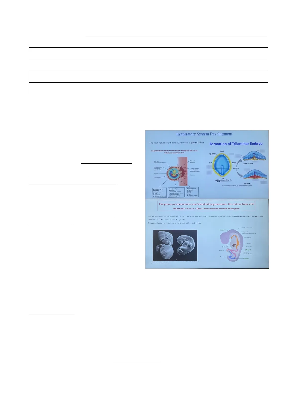
Dobbiamo parlare sia dello sviluppo del sistema respiratorio sia delle caratteristiche istologiche dei vari tratti che contraddistinguono questo Respiratory System Development sistema.
Eventi della Terza Settimana: Gastrulazione
The first major event of the 3rd week is gastrulation. Formation of Trilaminar Embryo Per capire meglio lo sviluppo del sistema respiratorio dobbiamo ricordare quelli che sono As gastrulation converts the bilaminar embryonic disc into a Head trilaminar embryonic disc. Cut edge of amnion gli eventi principali della terza settimana, in ined with endoderm) Maternal blood pool Left Righ (s) 14-15 days Sections particolare quegli eventi che portano alla Amniotic cavity containing Chorion | Served from blastocyst amniotic fuld trasformazione del disco bilaminare Amnion Adarbois f) Bilaminar embryonle Endometrium (h) 16 days dise dell'embrione in un disco trilaminare. L'evento Tall principale della terza settimana è infatti la Endoderm layer Mesoderm layer becomes ) Digestive system ) Circulatory system Lungs (epithelal layer) Skeletal system Ectoderm laye becomes gastrulazione, che ha inizio con la comparsa Pancreas 4) Lungs (inner layers) 4) Nervous System della linea primitiva che si forma in posizione caudale e sulla superficie dorsale The process of craniocaudal and lateral folding transforms the embryo from a flat embryonic disc to a three-dimensional human body plan. dell'epiblasto, nello specifico nella sua As a result of cephalocaudal growth and closure of the lateral body wall folds a continuously larger portion of the endodermal germ layer is incorporated into the body of the embryo to form the gut tube porzione mediale. Come conseguenza della The tube is divided into three regions: the foregut, midgut, and hindgut migrazione e della proliferazione delle cellule Primitive pharynx Foregut- Lung bud/ dell'epiblasto nella linea primitiva, si verrà Respiratory diverticulum Esophagus successivamente a formare il mesoderma, Liver - Pancreas Stomach ossia il foglietto che separerà epiblasto da Gallbladder Allantois- Midgut Primitive ipoblasto. Con la formazione della linea Intestinal loop Cloaca Hindgut primitiva si possono già definire gli assi di simmetria dell'embrione e in particolare l'asse cefalo-caudale, dorso-ventrale e destro-sinistro. Dopo la formazione di tutti e 3 i foglietti germinativi (ectoderma, mesoderma e endoderma), da cui poi origineranno tutti i tessuti del corpo umano, nella 4 settimana si verifica il ripiegamento dell'embrione non solo cefalo-caudale ma anche laterale.
Intestino Primitivo e Sviluppo Respiratorio
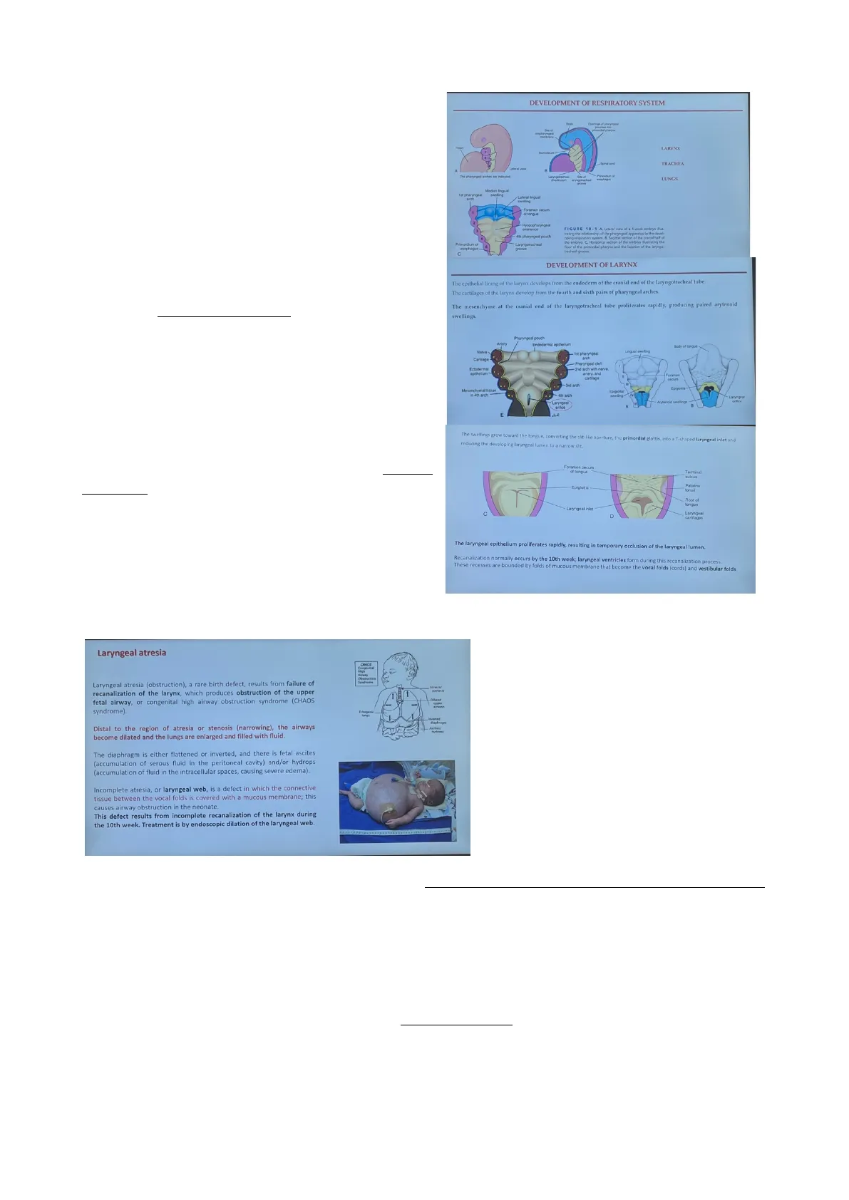
Come conseguenza di questo ripiegamento, l'endoderma verrà internalizzato e formerà il cosiddetto intestino primitivo. L'embrione, che fino ad ora era pressoché piatto, a questo punto è stato trasformato in una struttura tridimensionale. Questa ricapitolazione degli eventi della 3 e 4 settimana è importante ai fini dello studio del sistema respiratorio, poiché per il suo sviluppo è importante proprio l'intestino primitivo, che può essere suddiviso in una regione anteriore, detta intestino anteriore, e in un intestino medio e posteriore. Quello su cui bisogna focalizzarsi è l'intestino anteriore perché è proprio lì che si svilupperà il sistema respiratorio grazie all'interazione dell'endoderma con il mesoderma circostante (nello specifico il mesoderma splancnico). Infatti, nella diapositiva seguente si può notare che zoomando la parte dell'intestino primitivo, esso è caratterizzato dalla presenza di 5 tasche faringee: 1,2,3,4 e 6. La quinta tasca faringea non c'è perché non si forma proprio e se si forma regredisce molto velocemente. Daquesta struttura dell'intestino primitivo si formeranno la laringe, la trachea e i polmoni. In particolare, la laringe si forma come una piccola fessura tra la 4 e la 6 tasca faringea. L'epitelio di rivestimento della laringe, così come tutto il resto del rivestimento epiteliale, deriva proprio dall'endoderma di questa regione, mentre tutti i tessuti di supporto come la cartilagine e il muscolo, derivano dal mesoderma che circonda questa regione. Tra la 4 e la 6 tasca faringea si forma e sviluppa l'orifizio laringeo. Successivamente, grazie alla proliferazione delle cellule del mesoderma si generano due rigonfiamenti: uno da origine all'epiglottide, l'altro (quello inferiore) da origine alla laringe. Le cellule di questo rigonfiamento, in seguito a proliferazione, invadono l'orifizio laringeo (che ha la forma di una piccola fessura a T) andandolo a chiudere. La ricanalizzazione della laringe si verifica alla decima settimana con la formazione delle corde vocali e i ripiegamenti vestibolari. Ricordiamo che la laringe è coinvolta anche nella produzione dei suoni e della parola.
Sviluppo del Sistema Respiratorio
DEVELOPMENT OF RESPIRATORY SYSTEM Operings of pharyngeal primordial phanes oropharyngeal metran Heart LARYNX TRACHEA A The pharyngeal arches are indicated wyngotracheal esophagus LUNGS 1 st pharyngeal Lateral lingual arch swelling Foramen cecum if tongue Hyopopharyngeal eminence FIGURE 10-1 A, Lateral view of a 4-week embryo ilut- 4th pharyngeal pouch trating the relationship of the pharyngeal apparatus to the devel- oping respiratory system. B. Sagittal section Sag mal section of the cranial half of Primordium of esophagus Laryngotracheal oping respiratory ba embryo. C. Horin the embryo. C, Horizontal section of the embryo ilustraing the floor of the primordial pharynx and the location of the laryngo- tracheal groove C
Sviluppo della Laringe
DEVELOPMENT OF LARYNX The epithelial lining of the larynx develops from the endoderm of the cranial end of the laryngotracheal tube. The cartilages of the larynx develop from the fourth and sixth pairs of pharyngeal arches. The mesenchyme at the cranial end of the laryngotracheal tube proliferates rapidly, producing paired arytenoid swellings. Pharyngeal pouch Arten Endodermal epithelium Body of tongue- Nerve 1st pharyngeal Cartilage - Pharyngeal cleft Ectodermal and arch with nerve. epithelium artery. and Foramer cantlage cecum -3rd arch Epiglotis Epiglottal swelling' - Laryngeal Laryngen Arytenoid swelings office brfice E The swellings grow toward the tongue, converting the slit-like aperture, the primordial glottis, into a T-shaped laryngeal inlet and reducing the developing laryngeal lumen to a narrow slit. Foramon cocum of tongue Terminal sulcus Epiglottiş tons Root of tongue Laryngeal inlet Laryngoal D cartilages The laryngeal epithelium proliferates rapidly, resulting in temporary occlusion of the laryngeal lumen. Recanalization normally occurs by the 10th week; laryngeal ventricles form during this recanalization process. These recesses are bounded by folds of mucous membrane that become the vocal folds (cords) and vestibular folds.
Atresia Polmonare e Laringeal Web
ATRESIA POLMONARE E LARINGEAL WEB Laryngeal atresia CHAOS Congenital High Airway Obstruction Syndrome Atresia/ Ichogenk lungs Inverted diaphragm - Aurbes/ byťreps Ovviamente dal punto di vista clinico ci sono delle correlazioni che vanno ad interessare specificamente la laringe e una di queste è l'atresia laringea, ossia una malformazione congenita che prevede un'ostruzione della laringe dovuta ad una un mancanza e a difetto nella ricanalizzazione. Alla decima settimana, Incomplete atresia, or laryngeal web, is a defect in which the connective tissue between the vocal folds is covered with a mucous membrane; this causes airway obstruction in the neonate. infatti, la laringe è completamente occlusa This defect results from incomplete recanalization of the larynx during the 10th week. Treatment is by endoscopic dilation of the laryngeal web. dalle cellule epiteliali che hanno invaso l'orifizio laringeo, viene ricanalizzata e un difetto in questo processo porta ad atresia. L'occlusione delle vie aeree superiori causa una dilatazione del sistema respiratorio più profondo, come bronchi e polmoni, ed è caratterizzata anche dalla presenza di liquido nell'addome. Questa patologia può essere curata chirurgicamente non appena si nasce. Un'altra patologia è la laringeal web, che fa si che il tessuto connettivo (il tessuto di sostegno) sia ricoperto da una membrana mucosa e ciò ha come conseguenza un'ostruzione delle vie respiratorie.
Formazione di Trachea e Polmoni
Come si formano la trachea e i polmoni? Sempre nell' intestino anteriore, a partire dalla quarta settimana, si forma sulla sua superficie ventrale un diverticolo respiratorio, che nell'immagine è di colore azzurro. Questo diverticolo si forma grazie all'effetto dell'aumento di acido retinoico, che va a stimolare l'espressione del fattore di trascrizione TBX4 nell'endoderma. L'acido retinoico viene sintetizzato nel mesoderma e il suo effetto raggiunge l'endoderma: questo fa capire come l'interazione tra endoderma e mesoderma sia Laryngeal atresia (obstruction), a rare birth defect, results from failure of recanalization of the larynx, which produces obstruction of the upper fetal airway, or congenital high airway obstruction syndrome (CHAOS syndrome). Distal to the region of atresia or stenosis (narrowing), the airways become dilated and the lungs are enlarged and filled with fluid. The diaphragm is either flattened or inverted, and there is fetal ascites (accumulation of serous fluid in the peritoneal cavity) and/or hydrops (accumulation of fluid in the intracellular spaces, causing severe edema). Lateral view Median Ingual swelling Lingual swelling Mesenchymal tissue in 4th arch PalatinoFORMATION OF THE LUNG BUDS When the embryo is approximately 4 weeks old, the respiratory diverticulum (lung bud) appears as an outgrowth from the ventral wall of the foregut. Respiratory diverticulum Stomach Heart- Liver bud Duodenum Vitolina duc - Midgu Allantois Cloacal membrane Hence, epithelium of the internal lining of the larynx, trachea, and bronchi, as well as that of the lungs, is entirely of endodermal origin. The cartilaginous, muscular, and connective tissue components of the trachea and lungs are derived from splanchnic mesoderm surrounding the foregut. FORMATION OF THE TRACHEA This primordium of the tracheobronchial tree develops caudal to the fourth pair of pharyngeal pouches. Openings of pharyngeal pouches Primitive Pharynx Tracheorsephageal Trachromephagral seprom Opening of rimitive laryngotracheal divertirelem Laryngotracheal diverticulum Attachment of buccopharyngeal membrane bronchial bodd. Respiratory Sivertoulum B 4 weeks 4-5 weeks 5 weeks Laryngotracheal office When the diverticulum expands caudally, however, two longitudinal ridges, the tracheoesophageal ridges, separate it from the foregut. Subsequently, when these ridges fuse to form the tracheoesophageal septum, the foregut is divided into a dorsal portion, the esophagus, and a ventral portion, the trachea and lung buds. fondamentale per il corretto sviluppo del sistema respiratorio. L'espressione di specifici fattori a livello dell'endoderma e delle diverse strutture: ad esempio TBX4 è uno dei fattori fondamentali per la formazione del diverticolo respiratorio e sarà importante anche per l'allungamento e la diramazione del canale respiratorio, nello specifico per la formazione della trachea. Il rivestimento epiteliale di tutto il sistema respiratorio ha origine endodermica mentre il tessuto connettivo e la cartilagine, che ha un ruolo fondamentale nel sostenere e tenere aperto il canale respiratorio, hanno origine mesodermica. In corrispondenza tra la 4 e la 6 tasca faringea quindi si sviluppa il diverticolo respiratorio il cui lume è inizialmente collegato con il lume dell'esofago. Successivamente, due creste laterali vanno man mano a restringere il lume e successivamente a separarlo completamente, formando il setto tracheoesofageo. Esofago e intestino sono a questo punto separati: l'esofago si svilupperà posteriormente e la trachea anteriormente. La trachea verso la 4-5 settimana inizia ad allungarsi e contemporaneamente a biforcarsi e si formeranno due abbozzi bronchiali che si allungheranno progressivamente nella quinta settimana. È molto importante, per il corretto sviluppo del sistema respiratorio, l'espressione di alcune molecole che sono compartimentalizzate: prima ancora che si formi il setto tracheoesofageo c'è l'espressione di alcuni fattori come SOX e NBP7 nella parte dorsale, che sarà quella che poi darà origine all'esofago. Esattamente nella parte ventrale The regionalization of the different parts of the gut is controlled by the localized expression of different transcription factors. e nella parte che darà origine alla trachea invece vedremo l'espressione di NKX2, Notochord Blogg Dorsal Sh The unseparated anterior foregut tube shows high levels of Sox2, Noggin, and Noggin Bmp7 in the dorsal epithelium that will give rise to the esophagus. SHH(sonic hedgehog) e WNT6B. Nel Future esophagus Bmp7 The ventral epithelium, which will contribute to the trachea, highly expresses transcription factor Nkx2.1 and signaling molecules Shh and Wnt7b, along B with Rhou mesenchima fattori come Wnt2, wnt2b, fgf10 e bmp4 supportano l'espressione di Homeobox gene Barx1 is expressed at the demarcation between the dorsal and ventral foregut separation. questi geni nella componente epiteliale. The ventral mesenchyme factors Wnt2, Wnt2b, Fgf10, and Bmp4 support Future trachea gene expression in the epithelium. Quindi l'interazione tra mesenchima ed Defects in the Shh, Wnt, or Bmp pathway or mutations of Sox2, Nkx2.1, or Rhou can result in abnormal foregut development, leading to esophageal atresia with or without tracheoesophageal fistula. epitelio è fondamentale per il corretto Nkx2.1, Rhou me Shh, Wnt7b sviluppo del sistema e affinché esofago e Wnt2, Wnt2b Fgf10, Bmp4 Ventral trachea si separino correttamente. Schematic section showing dorsal-ventral patterning of the anterior foregut (mouse). Esistono anche in questo caso alcune correlazioni cliniche che vanno ad interessare specificamente la corretta separazione tra esofago e trachea: un esempio è l'atresia esofagea, in cui l'esofago non si forma in maniera corretta; molto spesso è associata ad una fistola tracheoesofagea, cioè l'esofago e la trachea che sono collegati in maniera scorretta.
Tipi di Atresia Esofagea
Esistono 4 tipi dii atresia esofagea:
The appearance and location of the lung bud are dependent upon an increase in retinoic acid (RA) produced by adjacent mesoderm. This increase in RA causes upregulation of the transcription factor TBX4 expressed in the endoderm of the gut tube at the site of the respiratory diverticulum. TBX4 induces formation of the bud and the continued growth and differentiation of the lungs Hindgu Initially, the lung bud is in open communication with the foregut. Separated pharyes bronchial bel
Non hai trovato quello che cercavi?
Esplora altri argomenti nella Algor library o crea direttamente i tuoi materiali con l’AI.
