Synaptic and Neuromuscular Transmission, Midwestern University
Slides from Midwestern University about Synaptic and Neuromuscular Transmission. The Pdf, a presentation for University students in Biology, details the mechanisms of neurotransmitter release and termination, and neural information integration, with explanatory diagrams.
Mostra di più14 pagine


Visualizza gratis il Pdf completo
Registrati per accedere all’intero documento e trasformarlo con l’AI.
Anteprima
Learning Objectives for Synaptic Transmission
- Be able to list and explain the major differences between electrical and chemical transmission.
- Be able to list and explain the major steps involved in chemical transmission.
- Explain the relationship between synaptic [Ca2+] and the amount of neurotransmitter released.
- Be able to explain the mechanisms for modulation of synaptic transmission under physiological conditions, and by drugs and disease.
- Be able to explain the steps in neuromuscular transmission and compare these steps to those in synaptic transmission between two neurons.
- Explain the mechanisms of action of various chemical agents and diseases that affect neuromuscular transmission.
Synapses Overview
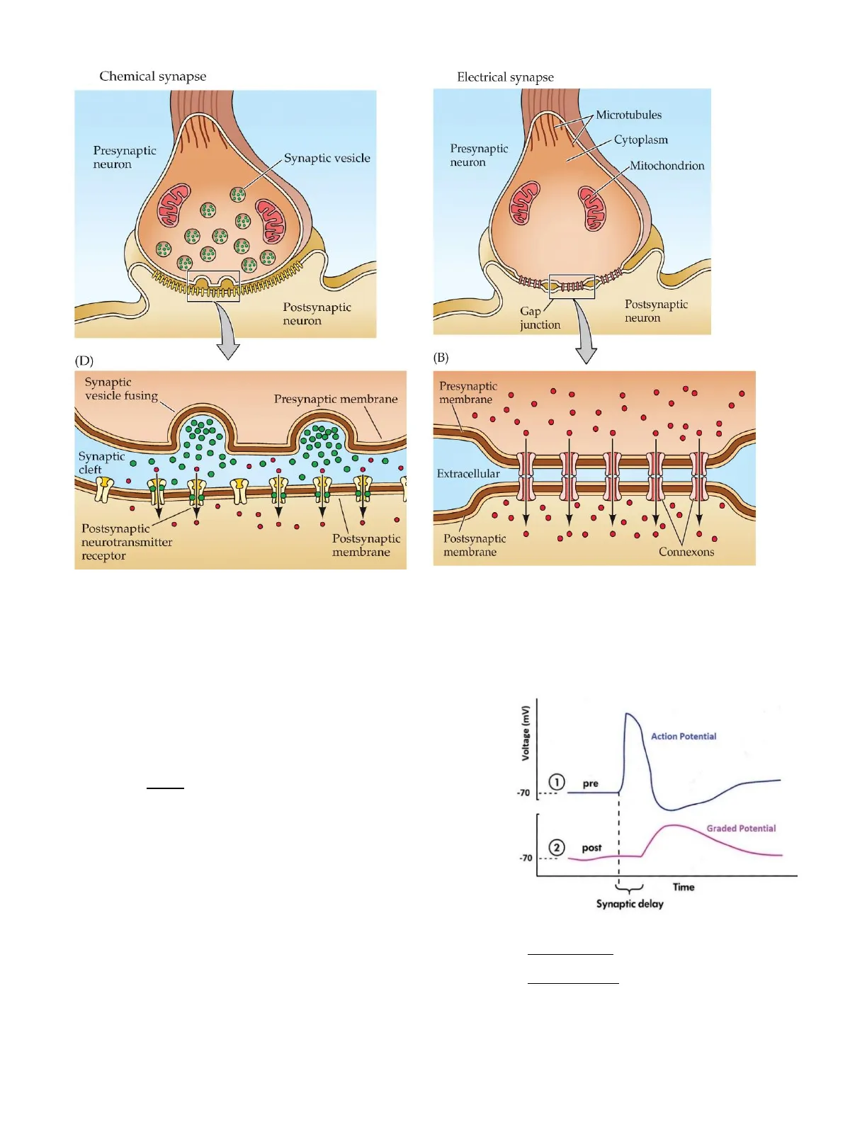
The region where an axon terminal meets its target cell is called a synapse (syn-, together + hapsis, to join). The neuron that delivers a signal to the synapse is the presynaptic cell, and the cell that receives the signal is called the postsynaptic cell. The narrow space between two cells is called the synaptic cleft. The synaptic cleft is filled with an extracellular matrix and the fibers hold the presynaptic and postsynaptic cells in position. There are two types of synapses: chemical and electrical synapses.
Chemical Synapses Characteristics
Most synapses in the body are chemical synapses where the presynaptic cell releases a chemical signal that diffuses across the synaptic cleft and binds to a membrane receptor on the postsynaptic cell.
Electrical Synapses Characteristics
Human CNS also contains electrical synapses that allow electrical current to pass through gap junction channels. Communication at electrical synapses is bidirectional and faster than at chemical synapses. Electrical synapses allow multiple CNS neurons to coordinate and fire simultaneously. They are not as common as chemical synapses and are largely concentrated in certain areas (e.g., the retina). Electrical synapses continually convey signals that can either excite or inhibit a coupled neighboring synapse, in contrast to neurotransmitter-based synapses, which typically exert either excitatory or inhibitory pulses of influence on the postsynaptic neuron.
Electrical and chemical synapses differ fundamentally in their transmission mechanisms.
Lecture #6 - Synaptic Transmission p. 6-1Chemical synapse Presynaptic neuron Synaptic vesicle PO Postsynaptic neuron (D) Synaptic vesicle fusing Presynaptic membrane Synaptic cleft Postsynaptic neurotransmitter receptor Postsynaptic membrane Fig. 1. Chemical synapse. At chemical synapses, there is no intercellular continuity, and thus no direct flow of current from the pre- to the postsynaptic cell. Synaptic current flows across the postsynaptic membrane only in response to the secretion of neurotransmitters, which open or close postsynaptic ion channels after binding to receptor molecules on the postsynaptic membrane.
Structure and Function of Chemical Synapses
There is a delay in transmission across the chemical synapse because that is the time necessary for the neurotransmitter to be released, diffuse across the synaptic cleft, and bind with receptors on the postsynaptic membrane (Fig. 4).
Electrical synapse Microtubules Cytoplasm Presynaptic neuron Mitochondrion Gap junction Postsynaptic neuron (B) Presynaptic membrane Extracellular Postsynaptic membrane Connexons Fig. 2. Electrical synapse. At electrical synapses, gap junctions occur between pre-and postsynaptic membranes. Gap junctions contain connexon channels that permit current to flow passively from the presynaptic cell to the postsynaptic cell.
Voltage (mV) Action Potential 1 pre -70 Graded Potential 2 post -70 Time Synaptic delay Fig. 4 Chemical synapse. Illustration of the action potential that might be recorded in the presynaptic cell and the delayed onset of a graded potential in the postsynaptic cell.
Lecture #6 - Synaptic Transmission p. 6-2
Structure and Function of Electrical Synapses
Although they are a distinct minority, electrical synapses are found in all nervous systems, including the human brain. Gap junctions consist of connexons, and hexameric complexes present in both the pre-and postsynaptic membranes. Channels assembled from connexons in these two membranes form pores that create electrical continuity between the two cells. The transmission between two neurons is extraordinarily fast and the communication can occur without the delay that is characteristic of chemical synapses (Fig. 5).
(A) (B) 25 Presynaptic neuron 0 -25 Membrane potential (mV) -25 -50 -50 Neuron 1 * * * * * * * 0 25 Postsynaptic neuron -25 0 -25 -50 -50 Minimal (<0.1 ms) synaptic delay 0 100 200 300 400 0 1 2 3 4 Time (ms) Time (ms) Fig. 5. The function of gap junctions at electrical synapses. (A) Rapid transmission of signals at an electrical synapse in the crayfish. An action potential in the presynaptic neuron causes the postsynaptic neuron to be depolarized within a fraction of a millisecond. (B) Electrical synapses allow the synchronization of electrical activity in hippocampal interneurons. The generation of an action potential in one neuron often results in the synchronized firing of an action potential in another neuron (asterisks).
Chemical Transmission Steps
Steps in Chemical Transmission Between Neurons
- Propagation of the action potential down the presynaptic axon results in depolarization of the membrane of the synaptic terminal.
- Voltage-gated calcium channels in the synaptic terminal open in response to depolarization. There are few, if any, Ca2+ channels elsewhere on neurons.
- Calcium enters the synaptic terminal through the open Ca2+ channels. Intracellular [Ca2+] is increased by the Ca2+ influx.
- Synaptic vesicles (containing neurotransmitters) fuse to the presynaptic membrane in a Ca2+- dependent fashion. Fusion of the vesicle membrane with the presynaptic membrane results in the rupture of the vesicle and the release of the neurotransmitter into the synaptic cleft (exocytosis).
- The neurotransmitter diffuses across the synaptic cleft and binds to specific receptors in the
postsynaptic membrane.
- Ion channels in the postsynaptic membrane open because of the binding of the neurotransmitter to the postsynaptic receptor.
- £ Local, graded potentials (postsynaptic potentials) occur because of the opening of postsynaptic channels. Either depolarize or hyperpolarize the postsynaptic membrane.
- Local current flow spreads to adjacent areas of the postsynaptic membrane. If the graded potentials are depolarizing and large enough, may elicit an action potential.
- The neurotransmitter is removed from the synaptic cleft and the membrane potential is restored to the previous level (i.e., the resting membrane potential).
1 An action potential depolarizes the axon terminal. 1 Synaptic vesicle with neurotransmitter molecules Action potential arrives at axon terminal. 2 The depolarization opens voltage- gated Ca2+ channels, and Ca2+ enters the cell. 3 Calcium entry triggers exocytosis of synaptic vesicle contents. 3 Ca2+ Docking protein 4 Neurotransmitter diffuses across the synaptic cleft and binds with receptors on the postsynaptic cell. - 2 4 5 Neurotransmitter binding initiates a response in the postsynaptic cell. Receptor Postsynaptic cell Voltage-gated Ca2+ channel 5 Cell response Fig. 6. Steps in neurotransmitter release.
Neurotransmitter Termination Mechanisms
Neurotransmitter action terminates when the chemicals are broken down, taken up into cells, or diffused away from the synapse (Fig. 7).
- Reuptake - the neurotransmitter is often taken up by the presynaptic cell for recycling.
- Diffusion - the neurotransmitter may just float out of the synaptic cleft.
- Degradation - enzymes are present in and around the synapse which degrade the neurotransmitter. Examples: catechol-O-methyltransferase (COMT) and monoamine oxidase (MAO) for sympathetic neurons and acetylcholinesterase for parasympathetic neurons.
The result of these processes is that the neurotransmitter signal is present for only a very short time (generally ms), in the absence of further stimulation.
Note that each of these steps is a common target for pharmacological intervention.
Lecture #6 - Synaptic Transmission p. 6-4 Synaptic cleftBlood vessel Axon terminal of presynaptic cell 1 Neurotransmitters can be returned to axon terminals for reuse or transported into glial cells. Synaptic vesicle 3 2 Enzymes inactivate neurotransmitters. Glial cell 3 Neurotransmitters can diffuse out of the synaptic cleft. 1 Enzyme Postsynaptic cell 2 Fig. 7. Neurotransmitter termination.
Integration of Neural Information
Divergence in Neural Communication
Divergence. Communication between neurons is not always a one-to-one event. Frequently, the axon of presynaptic neuron branches and its collaterals (branches) synapse on multiple target neurons. This pattern is known as divergence (Fig. 8).
Fig. 8. Divergence. In a divergent pathway, one presynaptic neuron branches to affect a larger number of postsynaptic neurons. Pearson education, Inc. 2019
Convergence in Neural Communication
Convergence. On the other hand, when a group of presynaptic neurons provides input to a smaller number of postsynaptic neurons, the pattern is known as convergence (Fig. 9).
Fig. 9. Convergence. In a convergent pathway, many presynaptic neurons provide input to influence a smaller number of postsynaptic neurons.
Spatial and Temporal Summation
A combination of convergence and divergence in the CNS may result in one postsynaptic neuron with synapses from as many as 10,000 presynaptic neurons (Fig. 10).
Lecture #6 - Synaptic Transmission p. 6-5dendrites cell body 0.1 mm dendrite Courtesy of Olaf Mundigl and Pietro de Camilli presynaptic nerve terminals (A) axon (B) Fig. 10. Thousands of synapses form on the cell body and dendrites of a motor neuron in the spinal cord. (A) Many thousands of nerve terminals synapse on this neuron, delivering signals from other parts of the animal to control the firing of action potentials along the neuron's axon. (B) A rat nerve cell in culture. Its cell body and dendrites (green) are stained with a fluorescent antibody that recognizes a cytoskeletal protein. Thousands of axon terminals (red) from other nerve cells (not visible) make synapses on the cell's surface; they are stained with a fluorescent antibody that recognizes a protein in synaptic vesicles, which are in the nerve terminals.
When two or more presynaptic neurons converge on the dendrites or cell body of a single postsynaptic cell, the response of the postsynaptic cell is determined by the summed input from the presynaptic neurons. The summed input from those synapses determines the activity of the postsynaptic neuron. The combination of several nearly simultaneous graded potentials is called spatial summation (Fig. 11). This refers to the fact that the graded potentials originate at different locations (spaces) on the neuron.
Spine head Spine neck Spines Excitatory synapses (red) Inhibitory synapses (blue) This illustration represents a three-dimensional reconstruction of dendritic spines and their synapses. Whether or not an action potential occurs in the postsynaptic cell depends on whether the summation of EPSPs and IPSPs results in reaching the threshold for the firing of APs or not (Fig. 12).
Fig. 11. Spatial summation. Multiple presynaptic neurons provide input on the dendrites and cell bodies of the postsynaptic neurons. This figure shows the dendritic spines of a postsynaptic neuron, with numerous excitatory and inhibitory synapses providing input. Pearson Education, Inc. 2019
Lecture #6 - Synaptic Transmission p. 6-6
Non hai trovato quello che cercavi?
Esplora altri argomenti nella Algor library o crea direttamente i tuoi materiali con l’AI.
