Embriologia della testa e della bocca: sviluppo dei denti umani, Università di Parma
Slide dall'Università di Parma su Embriologia della Testa e della Bocca: Sviluppo dei Denti Umani Odontogenesi. Il Pdf, una presentazione di Biologia a livello universitario, illustra la formazione di dentina, cemento e legamento parodontale, con illustrazioni dettagliate per una chiara comprensione dei processi biologici.
Mostra di più23 pagine

Visualizza gratis il Pdf completo
Registrati per accedere all’intero documento e trasformarlo con l’AI.
Anteprima
Apparato Stomatognatico: Morfologia e Funzione
DENTAL HYGIENE : MORPHOLOGY AND FUNCTION STOMATOGNATHIC APPARATUS
Embriologia della Testa e della Bocca
EMBRIOLOGY OF THE HEAD AND MOUTH: human teeth development
Odontogenesi
ODONTOGENESIS
ITAS UDIORUA CITAS . STUD UDIORU PARMENSIC UNIVERSITÀ DI PARMA Full Professor in Odontostomatologic Diseases Director European Master Degree in Oral Laser Applications (EMDOLA ) Director School of Speciality in Oral Surgery University of Parma UNIVERSITÀ DI PARMA EMDOLA European Master Degree in Oral Laser Applications
Organizzazione dei Denti
ODONTOGENESIS Teeth are organised into two opposing arches maxillary (upper) and mandibular (lower). These can be divided down the midline (mid-sagittal plane) into left and right halves. Teeth are positioned in alveolar sockets, and connected to the bone by a suspensory periodontal ligament.
Sviluppo dei Denti
- We shall look at the development of the teeth - their precursor tissues, differentiation, and related clinical conditions.
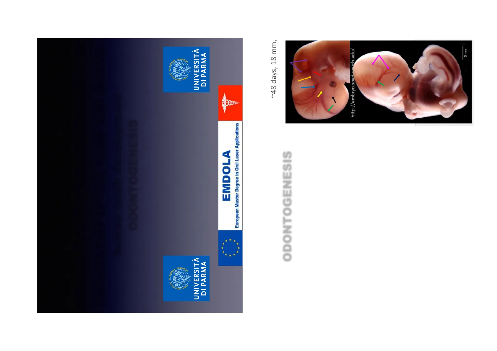
The first stage begins in the fetus at about 6 weeks of age. This is when the basic substance of the tooth forms. Next, the hard tissue that surrounds the teeth is formed, around 3 to 4 months of gestation. After the child is born, the next stage occurs when the tooth actually protrudes through the gum ~48 days, 18 mm, http://embryo.soad.umich.edu/ 2 mm
Prof. Paolo Vescovi DDS, PhD, MSc, Spec Oral Surgery MA · UNIVERSIT Dasser and · PARMENSISODONTOGENESIS Teeth develop and erupt at different times as a result of a complex, multi-step process, involving the patterning of inductive signals and homeobox genes. Signalling occurs between the oral epithelium and the underlying mesenchyme. Temporal and spatial control of gene expression leads to formation of different tooth types. Tooth development is recognised to occur in stages - initiation, morphogenesis, and differentiation.
Stadi di Sviluppo del Dente
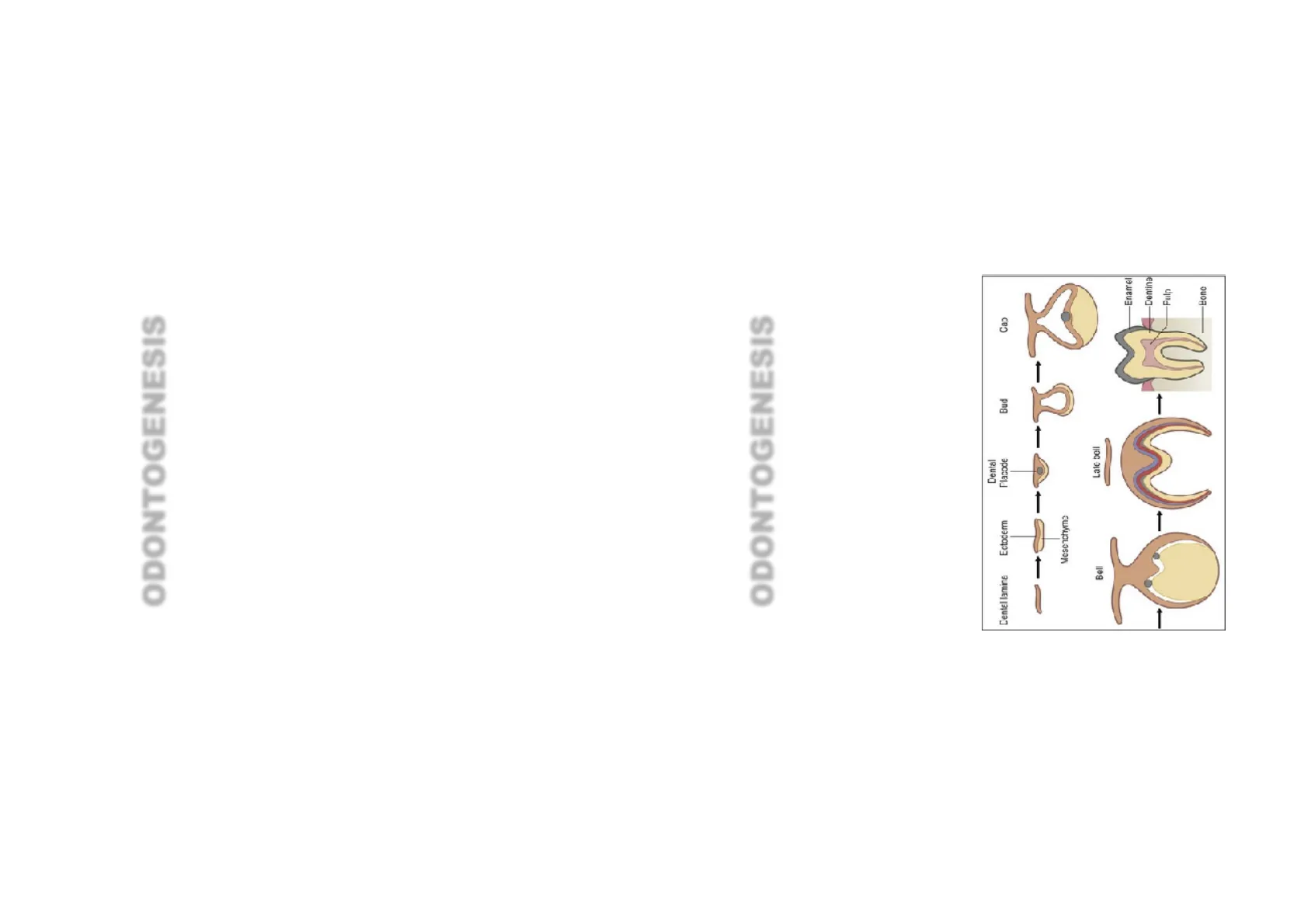
ODONTOGENESIS Tooth development is commonly divided into the following stages: the initiation stage, the bud stage, the cap stage, the bell stage, and finally maturation. The staging of tooth development is an attempt to categorize changes that take place along a continuum; frequently it is difficult to decide what stage should be assigned to a particular developing tooth. This determination is further complicated by the varying appearance of different histologic sections of the same developing tooth, which can appear to be different stages.
Dental Placode Dentel lamina Ectoderm Bud Cap - - + Mesenchyma Late bell Bell Enarrel Dentina - -Fulp -Bone
Odontogenesi: Stadio di Iniziazione
ODONTOGENESIS: INITIATION STAGE One of the earliest signs in the formation of a tooth that can be seen microscopically is the distinction between the vestibular lamina and the dental lamina. It occurs in the sixth to seventh week of the embryonic life. The dental lamina connects the developing tooth bud to the epithelial layer of the mouth for a significant time. This is regarded as the initiation stage.
Iniziazione del Dente
ODONTOGENESIS: INITIATION STAGE Tooth initiation begins at the 6th week in utero, when ectomesenchymal cells accumulate immediately below the oral epithelium. These cells are thought be derived from the neural crest cell population. The oral epithelium then proliferates down into the ectomesenchyme to form a primary epithelial band. At the 7th week in utero, the primary epithelial band differentiates into the vestibular and dental laminae:
- Vestibular lamina - forms the oral vestibule (the external opening to the oral cavity)
- Dental lamina - forms the teeth
Dental Placode Dental lamina Ectoderm Bud Cap + Mesenchyma Late bell Bell Enarrel Dentina - Fulp EoneDental lamina Ectoderm Dental Placode Bud Cap Mesenchyme Bell Late bell Enamel Dentine 1 1 Pulp Bone
Odontogenesi: Stadio a Gemma
ODONTOGENESIS: BUD STAGE The bud stage is characterized by the appearance of a tooth bud without a clear arrangement of cells. The stage technically begins once epithelial cells proliferate into the ectomesenchyme of the jaw. Typically, this occurs when the fetus is around 8 weeks old. The tooth bud itself is the group of cells at the periphery of the dental lamina. Along with the formation of the dental lamina, 10 round epithelial structures, each referred to as a bud, develop at the distal aspect of the dental lamina of each arch. These correspond to the 10 primary teeth of each dental arch, and they signify the bud stage of tooth development. Each bud is separated from the ectomesenchyme by a basement membrane. Ectomesenchymal cells congregate deep to the bud, forming a cluster of cells, which is the initiation of the condensation of the ectomesenchyme. The remaining ectomesenchymal cells are arranged in a more or less haphazardly uniform fashion.
Dental lamina Ectoderm Dental Placode Bud Mesenchyme
Odontogenesi: Stadio a Cappuccio
ODONTOGENESIS: CAP STAGE The first signs of an arrangement of cells in the tooth bud occur in the cap stage. A small group of ectomesenchymal cells stops producing extracellular substances, which results in an aggregation of these cells called the dental papilla. At this point, the tooth bud grows around the ectomesenchymal aggregation, taking on the appearance of a cap, and becomes the enamel (or dental) organ covering the dental papilla. A condensation of ectomesenchymal cells called the dental sac or follicle surrounds the enamel organ and limits the dental papilla. Eventually, the enamel organ will produce enamel, the dental papilla will produce dentin and pulp, and the dental sac will produce all the supporting structures of a tooth, the periodontiumEnamel Organ Dental Papilla
Odontogenesi: Stadio a Campana
ODONTOGENESIS: BELL STAGE The bell stage is known for the histodifferentiation and morphodifferentiation that takes place. The dental organ is bell- shaped during this stage, and the majority of its cells are called stellate reticulum because of their star-shaped appearance. The bell stage is divided into the early bell stage and the late bell stage. Cells on the periphery of the enamel organ separate into four important layers. Cuboidal cells on the periphery of the dental organ are known as outer enamel epithelium (OEE). The columnar cells of the enamel organ adjacent to the enamel papilla are known as inner enamel epithelium (IEE). The cells between the IEE and the stellate reticulum form a layer known as the stratum intermedium. The rim of the enamel organ where the outer and inner enamel epithelium join is called the cervical loop.
Odontogenesi: Organo dello Smalto
ODONTOGENESIS: ENAMEL ORGAN The enamel organ is composed of the outer enamel epithelium, inner enamel epithelium, stellate reticulum and stratum intermedium. These cells give rise to ameloblasts, which produce enamel and become a part of the reduced enamel epithelium (REE) after maturation of the enamel. The location where the outer enamel epithelium and inner enamel epithelium join is called the cervical loop. The growth of cervical loop cells into the deeper tissues forms Hertwig Epithelial Root Sheath, which determines the root shape of the tooth. During tooth development there are strong similarities between keratinization and amelogenesis. Keratin is also present in epithelial cells of tooth germ and a thin film of keratin is present on a recently erupted tooth (Nasmyth's membrane or enamel cuticle).
A C B Histologic slide showing a tooth bud. A: enamel organ B: dental papilla C: dental follicle
Odontogenesi: Papilla Dentale
ODONTOGENESIS: DENTAL PAPILLA The dental papilla contains cells that develop into odontoblasts, which are dentin-forming cells.Additionally, the junction between the dental papilla and inner enamel epithelium determines the crown shape of a tooth Mesenchymal cells within the dental papilla are responsible for formation of tooth pulp.
Enamel Organ Dental Papilla
Odontogenesi: Follicolo Dentale
ODONTOGENESIS: FOLLICLE C Histologic slide showing a tooth bud. A: enamel organ B: dental papilla C: dental follicle The dental sac or follicle gives rise to three important entities: cementoblasts, os teoblasts, and fibroblasts. Cementoblasts form the cementum of a tooth. Osteoblasts give rise to the alveolar bone around the roots of teeth. Fibroblasts are involved developing the periodontal ligament which connect teeth to the alveolar bone through cementum.
Morfogenesi Dentale
Morphogenesis Morphogenesis commences at the 10th week in utero, when five developing tooth germs appear in each quadrant, which give rise to the primary dentition. By the 16th week in utero, the tooth germs of the permanent incisors and the 1 st permanent molars begin to form. The 2nd and 3rd permanent molar tooth germs appear long after birth.
Denti Mascellari (Superiori)
Denti Primari Mascellari
Maxillary (upper) teeth Primary teeth
| Central incisor | Lateral incisor | Canine | First molar | Second molar | |
|---|---|---|---|---|---|
| Initial calcification | 14 wk I.U. | 16 wk I.U. | 17 wk I.U. | 15.5 wk I.U. | 19 wk I.U. |
| Crown completed | 1.5 mo | 2.5 mo | 9 mo | 6 mo | 11 mo |
| Root completed | 1.5 yr | 2 yr | 3.25 yr | 2.5 yr | 3 yr |
Denti Permanenti Mascellari
Maxillary (upper) teeth Permanent teeth
| Central incisor | Lateral incisor | Canine | First premolar | Second premolar | First molar | Second molar | Third molar | |
|---|---|---|---|---|---|---|---|---|
| Initial calcification | 3-4 mo | 10-12 mo | 4-5 mo | 1.5-2-2.25 yr | at birth | 2.5-3 yr | 7-9 yr | 1.75 yr |
| Grown completed | 4-5 yr | 4-5 yr | 6-7 yr | 5-6 yr | 6-7 yr | 2.5-3 yr | 7-8 yr | 12-16 yr |
| Root completed | 10 yr | 11 yr | 13-15 yr | 12-13 yr | 12-14 yr | 9-10 yr | 14-16 yr | 18-25 yr |
Denti Mandibolari (Inferiori)
Denti Primari Mandibolari
Mandibular (lower) teeth
| Initial calcification | Crown completed | Root completed | |
|---|---|---|---|
| Central incisor | 14 wk I.U. | 2.5 mo | 1.5 yr |
| Lateral incisor | 16 wk I.U. | 3 mo | 1.5 yr |
| Canine | 17 wk I.U. | 9 mo | 3.25 yr |
| First molar | 15.5 wk I.U. | 5.5 mo | 2.5 yr |
| Second molar | 18 wk I.U. | 10 mo | 3 yr |
Denti Permanenti Mandibolari
Mandibular (lower) teeth
| Central incisor | Lateral incisor | Canine | First premolar | Second premolar | First molar | Second molar | Third molar | |
|---|---|---|---|---|---|---|---|---|
| Initial calcification | 3-4 mo | 3-4 mo | 4-5 mo | 1.5-2 yr | 2.25-2.5 yr | at birth | 2.5-3 yr | 8-10 yr |
| Crown completed | 4-5 yr | 4-5 yr | 6-7 yr | 5-6 yr | 6-7 yr | 2.5-3 yr | 7-8 yr | 12-16 yr |
| Root completed | 9 yr | 10 yr | 12-14 yr | 12-13 yr | 13-14 yr | 9-10 yr | 14-15 yr | 18-25 yr |
Morfogenesi della Corona Dentale
ODONTOGENESIS: MORPHOGENESIS The crown of the tooth, which is influenced by the shape of the inner enamel epithelium, also takes shape during this stage. Throughout the mouth, all teeth undergo this same process; it is still uncertain why teeth form various crown shapes-for instance, incisors versus canines. There are two dominant hypotheses. The "field model" proposes there are components for each type of tooth shape found in the ectomesenchyme during tooth development. The components for particular types of teeth, such as incisors, are localized in one area and dissipate rapidly in different parts of the mouth. Thus, for example, the "incisor field" has factors that develop teeth into incisor shape, and this field is concentrated in the central incisor area, but decreases rapidly in the canine area.
Non hai trovato quello che cercavi?
Esplora altri argomenti nella Algor library o crea direttamente i tuoi materiali con l’AI.
