Clinical Anatomy: Clinical Pathologies and Diagnostic Imaging of the Lungs
Slides from Medready.org about Clinical Anatomy. The Pdf discusses clinical pathologies and diagnostic imaging of the lungs, covering topics like atelectasis, pneumothorax, and pleural effusion. The Pdf is a valuable resource for university students studying Biology.
See more67 Pages

Unlock the full PDF for free
Sign up to get full access to the document and start transforming it with AI.
Preview
Clinical Anatomy Lecture: Clinical Pathologies and Images of the Lungs
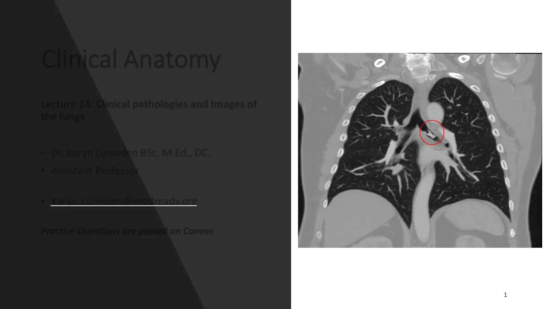
· Dr. Karyn Lumsden BSc, M.Ed., DC. · Assistant Professor · Karyn.Lumsden@medready.org Practice Questions are posted on Canvas Can you spot the problem? T C 0 0 0 0 0 0 0 1Learning Objectives:
- Discuss clinical pathologies related to the respiratory system and understand their pathogenesis, clinical symptoms, diagnosis, and treatment
- Pulmonary collapse: Atelectasis & pneumothorax
- Pneumothorax: Closed, Open and Tension
- Pleural effusion: different types of fluid collection
- Pneumonia: infiltration of different lobes
- Pulmonary embolism
- Define and discuss treatment strategies with respect to lung diseases
- Bronchoscopy
- Chest tube thoracostomy
- Thoracentesis
Recall: Anatomy of the Lungs
Superior - Right upper lobe - RUL Middle - Right middle lobe - RML Inferior - Right lower lobe - RLL RIGHT LUNG LEFT LUNG SUPERIOR (UPPER) LOBE @ 123RF SUPERIOR (UPPER) LOBE Superior - Left upper lobe - LUL Inferior - Left lower lobe - LLL MIDDLE LOBE INFERIOR (LOWER) LOBE 23RF INFERIOR (LOWER) LOBE
Auscultation of the Lungs
- Best heard with a stethoscope
- Normal lung sounds occur in all parts of the chest area, including above the collarbones and at the bottom of the rib cage. Normal sounds of Auscultation: Sound Quality Location Vesicular Soft/ low pitch Both lung fields Bronchial- vesicular Intermittent intensity 1st and 2nd intercostal space Bronchial Soft Over sternum Tracheal Loud and high pitched Neck 1 -2 1 2 4 3 5 6 4 + 3 B 8 7 5 6 10 8 - 72 1 Y 3 4 5 6 8 7 9 10 1-2 4 + 3 5 - 6 8 7 Auscultation of the Lungs
- Absent or decreased sounds can mean:
- Air or fluid in or around the lungs
- Increased thickness of the chest wall
- Over-inflation of a part of the lungs
- Reduced airflow to part of the lungsAuscultation of the Lungs
- The 4 most common abnormal breath sounds are:
- Rales = Small clicking, bubbling, or rattling sounds in the lungs. They are heard when a person inhales/inspires. Result of fluid build up in smaller airways
- Pneumonia, Pleural effusion
- Rhonchi = Low pitched, clunky or rattling sounds (resembles snoring) most pronounced with person inhales/inspires. Usually improve when person coughs / expels mucus
- Chronic Obstructive Pulmonary disorder (COPD), Cystic Fibrosis (CF), Chronic bronchitis
- Stridor = Harsh, vibratory sound heard when a person inhales/inspires. Usually, it is due to a blockage/narrowing in the upper airway
- Foreign body aspiration (trachea)
- Epiglottitis, Narrowed voice box
- Wheezing = High-pitched whistling sounds produced by narrowed airways. Heard most often during exhalation AND inhalation and can be heard without the use of stethoscope.
- Asthma
Clinical Correlation: Aspiration of a Foreign body
- Object inhaled into the airways
- Location depends on size of object and position of patient
- 30-40% end up in the Right primary (main) bronchus (m)
- 5-15% up in in the Right secondary inferior lobar bronchus (i)
- If the object is small enough (gravity pulls it down!)
- Tertiary (segmental) bronchi
- If upright - posterior basal segment of inferior lobe
- If supine - superior segment of inferior lobe M I S P 81Clinical Correlation: Aspiration of a Foreign body
- Usually occurs in young children
- In adults, rare or by accident
- Symptoms:
- Sudden onset dyspnea (shortness of breath)
- Cough, wheezing, stridor
- Decreased breath sounds
- Gagging
- Drooling L AP XRAY BRONCHOSCOPY
Treatment: Bronchoscopy
- Bronchoscope - thin tube passed through nose or mouth, down the throat and into the lungs
- Endoscopic camera for visualizing the bronchial tree
- Can be used to:
- Obtain samples of mucus or tissue
- Remove foreign bodies or other blockages
- Provide treatments - stent placement for example 18/15/201 PADRE CRISE BRONCHOSCOPY Facility SONY Mercy if O e Ages D. 07 10 CT: 15/2015 10:50 Phys Comment: IMAGE HDClinical Correlation: Aspiration of a Foreign body
- Aspiration of a foreign body in older adults = contrast media into bronchial tree
- Normally barium is used with X-ray examination of GI system
- Barium aspiration typically seen in elderly/adults who suffer from dysphagia
- Treatment involves: 100% oxygen, lung wash with NaCL to flush out barium or vibration therapy to help patient remove/cough out foreign material AP Xray
POLLING QUESTION 1: Foreign Body Aspiration Pathway
A 2-year-old boy was brought to the emergency department after his older brother informed their parents that he shoved a bead up his nose while they were playing. The physician is concerned about an aspirated body. Which is the most likely pathway of the bead in this particular case? A. Right primary bronchus > right inferior lobar bronchus > posterior basal segment B. Left primary bronchus > left inferior lobar bronchus > posterior basal segment C. Right primary bronchus > right inferior lobar bronchus -> superior segment D. Left primary bronchus > left inferior lobar bronchus -> superior segment E. Right primary bronchus > right inferior lobar bronchus -> inferior segment F. Left primary bronchus > left inferior lobar bronchus > inferior segment
Lung Tissue Pathologies Overview
- Pulmonary collapse - buildup of pressure inside or around the lungs causing all or some of the lung to collapse
- Pressure build up can be caused by air or fluid accumulation
- Pleural effusion - buildup of excess fluid in pleural cavity
- Pneumonia - respiratory infection characterized by inflammation of the alveolar space and/or interstitial tissue of the lungs
- Pulmonary embolism (PE) - obstruction of the pulmonary artery and/or one of its branches by a thrombus, usually from the deep vein system of the legs or pelvis AP XRAY A
Lung Tissue Pathologies: Pulmonary Collapse
Pulmonary Collapse = buildup of pressure inside airways or within pleural cavity causing all or some of the lung to collapse
- Pressure build up can be caused by air or fluid accumulation
- Symptoms:
- shortness of breath
- chest pain, which may be more severe on one side of the chest
- sharp pain when inhaling
- pressure in the chest that gets worse over time
- Cyanosis = blue discoloration of the skin or lips
- increased heart rate
- rapid breathing
- confusion or dizziness Diagnosis
- Patient history to determine cause
- Chest X-ray
- How to determine pulmonary collapse on X-ray . Linear shadow of visceral pleura with lack of lung markings peripheral to the shadow may be observed . Look at spaces between the ribs to see if there's any tissue present
Lung Tissue Pathologies: Causes of Pulmonary Collapse
Causes of Pulmonary Collapse: ATELECTASIS = alveolar sacs cannot inflate properly because of a blockage or pressure inside the airways PNEUMOTHORAX = excess air, or fluids, are stuck in the pleural cavity, causing lung to collapse Blockage of airways leading to buildup of pressure inside lungs and/or airways Buildup of pressure outside lungs in thoracic cavity (air outside of lungs) Causes: Foreign body aspiration Tumor Mucus plug Mucous plugs accumulating B Mucous plug Air absorbed from alveoli; lung segment collapses A Alveoli lined by flattened epithelium to allow gas exchange C Causes: Disruption/puncture to pleura Trauma to chest Spontaneous Trachea Collapsed lung Normal lung Air or fluid in the pleural space Look for one of these causes in the question stem to help differentiate !!
Lung Tissue Pathologies: Tracheal Deviation
- Pleural pressures on either side of the trachea determine its position within the mediastinum.
- Changes in pressure will lead to noticeable deviation "shift" of the trachea which is detectable on x-ray
- The trachea will shift towards the side with relatively higher negative pressure compared to the opposite side. Pneumothorax Left lung Inspiration Diaphragm Trachea pushed away from injured side = Pneumothorax or Pleural effusion Atelectasis NL Trachea pulled towards injured side = AtelectasisTrachea deviated towards diseased side = Atelectasis Trachea deviated away from diseased side = Pneumothorax or Pleural effusion A.P. SITTING AP XRAY
Clinical Correlation: Atelectasis
- Sudden onset of a partial or complete collapsed lung
- Blockage of airways leading to buildup of pressure inside lungs and/or airways Atelectasis Obstruction in bronchus Airflow obstructed Remaining air diffuses into tissues and is not replaced Nonaeration and collapse Diagnosis Patient history may include one of the key causes:
- Foreign body aspiration
- Tumor
- Mucus plug Imaging:
- Signs of pulmonary collapse
- Evidence of foreign body in airway
- Tracheal deviation towards side of blockage
Clinical Correlation: Atelectasis Patient History
Patient History of present illness: . A 10-month-old male presented after choking on a carrot. Mom reported coughing with perioral cyanosis. · Auscultation revealed stridor in the right lung fields particularly when coughing. Significant findings: · Chest radiograph showed increased radiolucency (red arrow) and flattening of the diaphragm on the right side (blue arrow), as well as left mediastinal shift (green arrow), indicating obstruction. · Airway deviation? L AP Upright PA Increased radiolucency Left mediastinal shift Flatten right diaphragm
Clinical Correlation: Pneumothorax Types
Pneumothorax Closed pneumothorax Open pneumothorax Tension pneumothorax Air in pleural space Air in pleural space Air in pleural space increasing and unable to escape https://youtu.be/B3-EULlh7_s
Clinical Correlation: Closed / Spontaneous / Non-traumatic Pneumothorax
Closed / Spontaneous / Non-traumatic Pneumothorax
- Sudden onset of a collapsed lung without any apparent cause
- Spontaneous communication between the alveolar spaces and pleural space
- Primary Spontaneous = idiopathic (No known trauma)
- Males, tall thin stature at slightly higher risk
- Secondary Spontaneous = can occur secondary to a variety of lung diseases and disorders (COPD, CF, severe asthma, etc.)
- Look for any of these causes in the question stem to help with diagnosis Air in pleural space 0 0 0 0 0 Closed pneumothorax
Clinical Correlation: Spontaneous Pneumothorax Diagnostic Findings
Clinical Correlation: Spontaneous Pneumothorax Diagnostic findings: . Patient History: · No obvious signs of trauma given in question or seen in imaging · Possible history of asthma, COPD, bronchitis
- Imaging characteristics: . Increased lucency of intercostal spaces in left thoracic cavity R BMM 1/2 AP XRAY Can you see the tracheal deviation?
Can’t find what you’re looking for?
Explore more topics in the Algor library or create your own materials with AI.
