Lecture on vascular physiology, blood composition, and vessels
Slides from University about Lecture 13: Vascular Physiology. The Pdf, a presentation for university-level Biology students, delves into cardiovascular physiology, detailing blood components and vessel types. It also covers Starling forces regulating fluid movement across capillary walls.
See more11 Pages

Unlock the full PDF for free
Sign up to get full access to the document and start transforming it with AI.
Preview
Cardiovascular Physiology Overview
Learning Objectives for Vascular Physiology
- Name and describe the components of blood.
- Characterize and differentiate between the various blood vessels.
- Describe the regulation of blood flow and factors that influence blood flow.
- Differentiate between turbulent and laminar flow.
- Define MAP and factors that determine MAP.
- Contrast the magnitude of blood pressure, resistance, cross-sectional area, and velocity of blood flow through the cardiovascular (pulmonary and systemic) system.
- List five factors that determine fluid movement across the capillary wall.
- Describe how changes in vascular resistance and protein concentration impact fluid movement.
Blood Components
Components of Blood: Plasma and Formed Elements
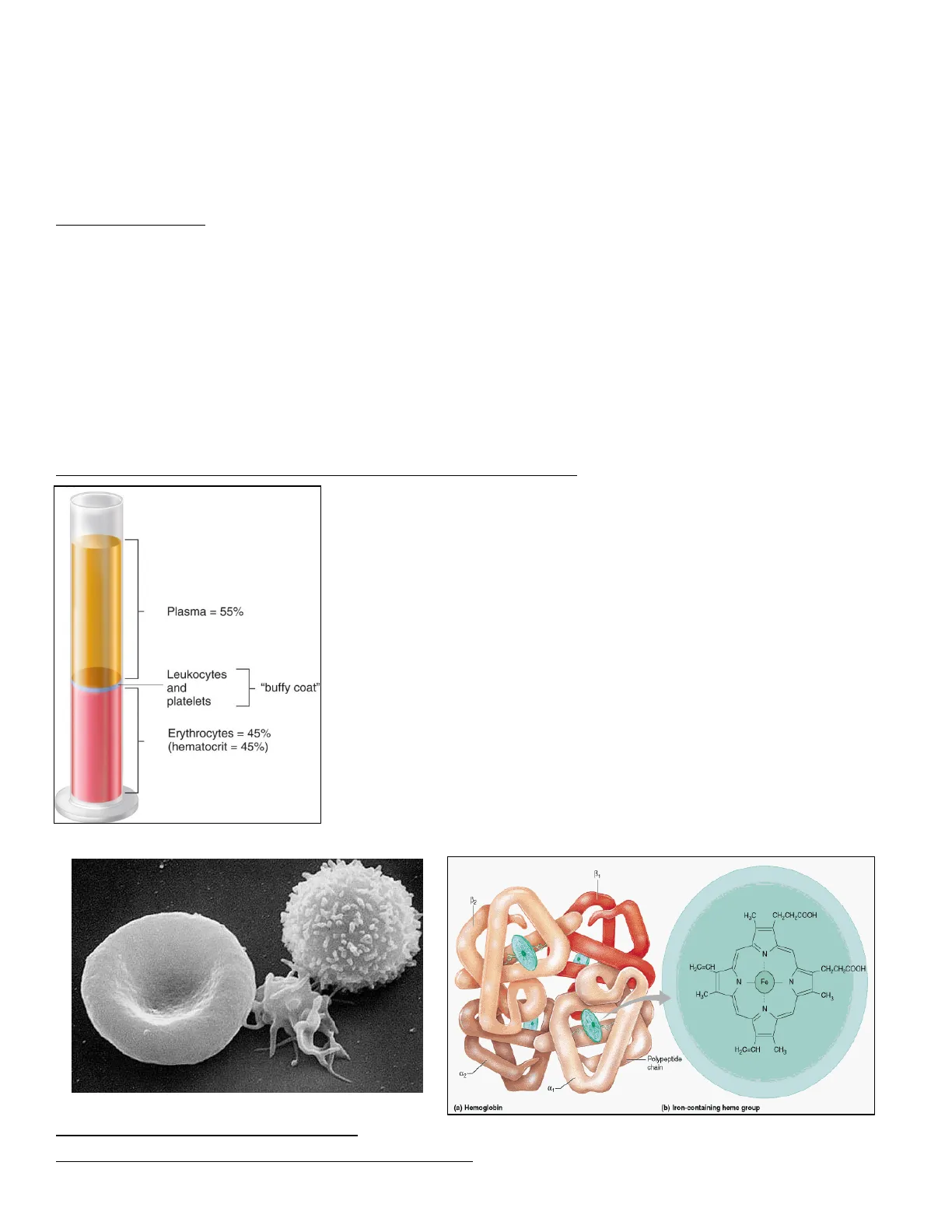
Blood is composed of plasma and formed elements (Fig 1). Plasma comprises about 55% of blood volume and contains electrolytes, proteins, gases, clotting factors, hormones, and glucose. Serum is the portion of plasma that does not contain clotting factors. Formed elements comprise the remaining 45% of blood and include erythrocytes, lymphocytes, and platelets (Fig 2).
In cardiovascular physiology, the primary focus will be on erythrocytes of the three types of formed elements. These cells contain an iron (Fe)- containing protein called hemoglobin (Hb), about 280 million per erythrocyte (Fig 3). Each of the iron atoms binds one molecular oxygen (O2).
Blood Vessels Characterization
Characterizing Different Blood Vessels
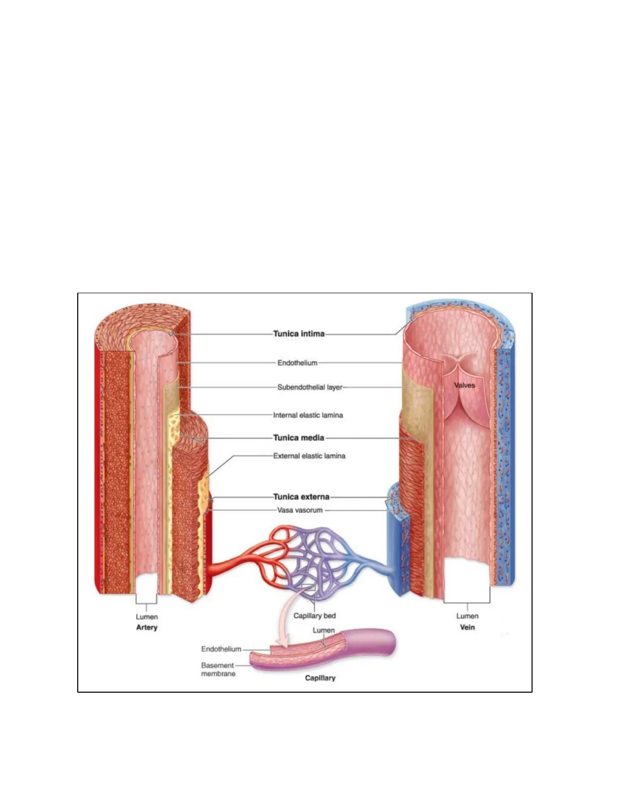
Arteries are known as "conduit vessels". These vessels have low and unchanging resistance to flow. They are characterized by endothelium, thick smooth muscle, elastic connective laminar tissue, and a smaller lumen compared to veins.
Arterioles are known as "resistance vessels". These vessels have more smooth muscles than arteries and therefore thicker walls. However, arterioles are less elastic than arteries.
Capillaries are known as "exchange vessels". These vessels are marked by an endothelial layer and have the smallest diameter of all the vessels. The thin vascular wall allows for exchange of substances, including glucose, gases, and ions.
Veins and venules are known as "capacitance vessels". These vessels have a thin smooth muscle wall and are therefore distensible. Veins and venules have the largest diameter of all vessels and carry more than 50% of all systemic blood. These vessels also have valves that prevent backflow of blood.
Circulatory System Organization
The vessels are organized into a circulatory system that transports blood throughout the body (Fig 5). The two branches of the circulatory system are the systemic and pulmonary.
These two circulations are in series that is, all the blood that flows through the systemic circulation must flow through the pulmonary; the output of the left ventricle equals the output of the right ventricle.
Blood Flow Regulation
Regulation and Factors Influencing Blood Flow
Blood flow is highly regulated so that appropriate proportions of ventricular output go to various organs for its proper functioning (Fig 6). Regulation of blood flow assures blood supply to vital organs of the body and coordinates cardiac output with metabolic needs of the body.
Flow (Q) through a tube is defined as: Q=AP/R
Flow through the cardiovascular system or through a single blood vessel is dependent on two things:
- The pressure difference (AP) at any two points along the blood vessel.
- Flow only occurs if AP exists.
- Resistance (R) changes can cause flow to change. Ex: At the same AP, high R has lower Q than low R (Fig 7).
- The resistance (R) to flow produced by the geometry of the vessel. R = 8ηl/πι4
Vessel Resistance Factors
Vessel resistance is dependent upon:
- The viscosity (n) of the fluid that is flowing
- The length (I) of the vessel
- The 4th power of the radius (r4)of the vessel
Given that viscosity and length are not easily manipulated, the radius of the vessel has the greatest influence on R. The vessel radius is regulated by changes in sympathetic nerve activity to the smooth muscle in the vessel wall.
The formula for R can be substituted in the flow formula to give the Poiseuille Equation:
ΔΡ πι4 Q= 8nl
Radius and Pressure Relationship with Resistance
Important points to note about the relationship of radius and pressure with resistance:
Resistance and vessel radius - The radius of a vessel is inversely related to the resistance of the vessel (as radius decreases, resistance increases); radius has a powerful effect on resistance because resistance changes as the 4th power of radius; this means that if the radius decreases by 1/2, the resistance increases 16 times.
When calculating resistance, the effect of resistance on pressure depends upon how the resistance is organized (Fig 8).
- When resistors are arranged in series, resistances are summated.
- When resistors are arranged in parallel, resistance adds as the reciprocals. The total resistance of parallel arrangement is less than any single resistance.
Turbulent vs. Laminar Flow
Differentiating Blood Flow Types
Laminar flow (Fig 9a) is the normal, streamlined, longitudinal flow of blood travelling in parallel through a vessel. The lowest velocity of blood is towards the vessel wall, while the highest velocity of blood is in the middle of the vessel.
Turbulent flow (Fig 9b) is the nonlinear, rapid, irregular flow of blood which is often seen in atherosclerosis (Fig 10), stenotic arteries, and stenotic heart valves (Fig 11).
Turbulent Flow in Blood Pressure Measurement
Turbulent flow may also be created and heard during blood pressure measurements using a sphygmomanometer (Fig 12). A cuff is placed around the upper arm with a stethoscope held under the base of the cuff just above the crease of the elbow. With the valve of the pump closed, the cuff is inflated until the brachial artery is occluded. At this point, there is no blood flow through the brachial artery and no sound heard through the stethoscope. As the pressure of the cuff is released slowly, the brachial artery is partially occluded allowing turbulent flow of blood through the vessel and Sounds of Korotkoff are heard. Once the pressure off the brachial artery is released and the brachial artery is completely opened, blood returns to laminar flow and no sound is heard through the stethoscope.
Mean Arterial Pressure (MAP)
Defining MAP and Its Determinants
The first Sound of Korotkoff heard through the stethoscope is the systolic blood pressure. The last Sound of Korotkoff heard through the stethoscope is the diastolic blood pressure (Fig 13).
The maximal aortic pressure created by ventricular ejection of blood during systole is the systolic blood pressure. Systolic pressure reflects cardiac output and the normal value is 120 mmHg.
The minimum aortic pressure created by ventricular filling of blood during diastole is the diastolic blood pressure (Fig 14). Diastolic pressure reflects the resistance of the vessels and the normal value is 80 mmHg.
Systolic and diastolic pressure values are often expressed as 120/80. Pulmonary blood pressures are notably lower. The difference between systolic and diastolic pressures is pulse pressure.
Blood pressure is pulsatile (Fig 13). The magnitude of pressure and its pulsatile nature decreases from arterial side to venous side of the circulation.
Mean arterial pressure (MAP) may be calculated from the systolic and diastolic pressures:
MAP = Diastolic + 1/3(Pulse Pressure)
If normal systemic blood pressure is 120/80, MAP equals 93 mmHg.
Blood Flow Dynamics Across Systems
Blood Pressure, Resistance, Area, and Velocity in Cardiovascular System
Figure 15 and 16 illustrates the various differences amongst vessels.
Arteries have the highest systolic and diastolic pressures of all the vasculature. These vessels have the greatest velocity of blood flow, but low cross-sectional area.
The arterioles have the greatest relative resistance (RR) due to strong regulation by the sympathetic nervous system.
Capillaries are vessels of smallest diameter but because of their high number and parallel arrangement, their resistance is lower than that of arterioles. The high number and parallel arrangement also makes them have the greatest cross-sectional area. Due to their large cross-sectional area velocity of blood flow is at its lowest enabling exchange of materials between blood and tissue.
As capillaries coalesce into venules and veins, velocity of blood flow increases and cross-sectional area decreases. Veins are very compliant and so they hold a large volume at low pressure. Because of this most of the blood volume is on the venous side of the systemic circulation. In a subsequent lecture we will see that contracting venous smooth muscle decreases venous compliance and the blood volume of the veins. This helps bring blood toward the heart during times of reduced cardiac output.
In the pulmonary circulation, blood pressures are lower than in the systemic circulation. The capillaries are still the site of highest cross-sectional area and lowest flow velocity.
Capillary Fluid Movement
Factors Determining Fluid Movement Across Capillary Wall
Learning Objective #7: List five factors that determine fluid movement across the capillary wall.
Can’t find what you’re looking for?
Explore more topics in the Algor library or create your own materials with AI.
