Radiology of the Abdomen: Indications, Patient Preparation, and Techniques
Slides about Radiology of the Abdomen. The Pdf, a university-level presentation, illustrates abdominal radiology, including indications, patient preparation, and standard and additional radiographic techniques. It presents clinical cases with radiographic images of masses and foreign bodies.
See more24 Pages

Unlock the full PDF for free
Sign up to get full access to the document and start transforming it with AI.
Preview
Radiology of the Abdomen: Indications
Radiology of the abdomen -it is indicated for a wide variety of condition:
- gastrointestinal disease:
- vomiting
- suspected foreing body
- tenesmus
- diarrhoea
- urogenital disease:
- dysuria, stranguria
- haematuria
- urinary incontinence
- anuria, polyuria
- non-specific disease:
- abdominal distension
- palpable abdominal masses
- or organomegaly
- abdominal pain
- pyrexia of unknown origin
- weight loss
- screening for primary or secondary neoplasia
- screening for abdominal injury following trauma
Patient Preparation for Abdominal Radiography
- it is recommended that food is withheld for 12 hours to ensure that the stomach and small intestine are relatively empty;
- the patient should be given the opportunity to defecate and urinate prior to abdominal radiography;
Standard Radiographic Views
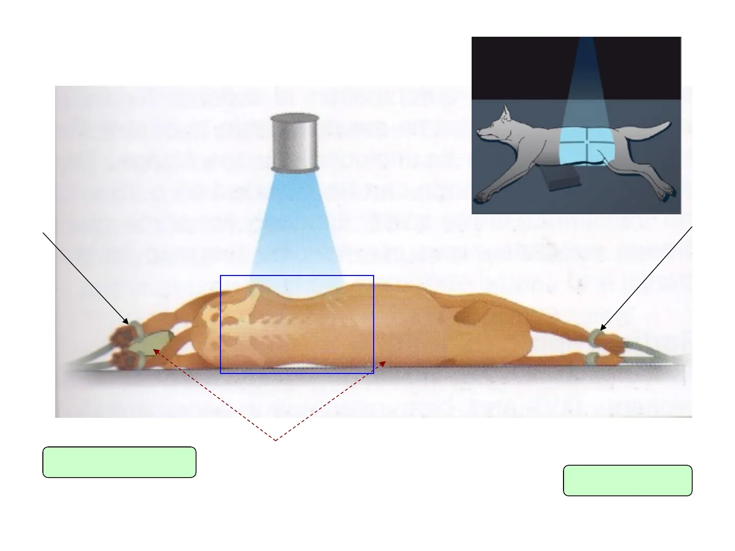
Standard radiographic views: -LL + VD = minimum recommendation; -in cases of suspected gastrointestinal obstruction = L left + L right + VD (as this allows redistribution of gas and fluid within the intestin and stomach)
Additional Radiographic Views
Additional views: -DV (as part of a barium gastrogram); -lateral oblique views (to evaluated the terminal ureters when performing iv urography); -horizontal beam decubitus view (to detect small volumes of free abdominal gas, which typically collects at the higest point of the abdominal beneath the non- dependent ribs with the animal positioned in lateral recumbency);-cats + small dogs -large dogsI- - -Acq st Gb Bd Pd B APd LL S RK Duo RL LK
Pancreas Visibility on Radiography
-the normal pancreas is too small to be visible on radiography, so any displacement due to changes in adjacent organs will not be recognized; -ultrasonography or CT are more sensitive;ABDOMEN Mag: 1.0x 10 2 4 11 7 6 8 13 12 1 3 9 5 R westie , 10 ani
Abdominal Organ Identification
1-liver, 2-stomach fundus region, 3-stomach pylorus region, 4-spleen head, 5- spleen tail, 6-duodenum, 7-colon, 8-cecum, 9-small intestines, 10-right kidney, 11- left kidneys, 12-colon, 13- urinary bladder.1-stomach fundus region, 2-stomach pylorus region, 3-spleen head 4-cecum 5-left kidney 6-colon 7-transvers colon 8-liver 9-duodenum 10-urinary bladder va01 Facultatea de Medicina Vet AV7QXPO.1 L O Basmatra 8 2012 Acq Tm: 1 IEN 9 Dx 2 1 7 3 5 4 6 10 M / Lin: DCM / Id: ID L:2047Right kidney Stomach Left kidney Colon Liver Urinary bladder Cecum Colon Tail of the spleenpekinez 3 ani M Lin: DCM / Lin: DCM / Id: ID VALA0OF 1.00.17 01 Le Medicina Veterinara B 4FQX2.1 O 20090417- ape 2009 Acq Tm: pekinez M 3 R in: DCM / Id: ID 2047
Normal X-ray with Contrast Medium
Normal x-ray with contrast mediumABDOMEN ZUT Acq Tm: L Normal x-ray with contrast medium R 1.2 ani M DOM / INIx x x x x x X - Dark gray - fat, that gives contrast for the other organs in the abdominal cavity X - Fecalomas on the colonA 2013 Feb Acq Tm: 13:00 N DOMEN imediat dupa administra℮ :DCM / Lin: DCM / |/ Lin: DCM / Id: ID :2047 SIZES ARE APPROXIMAR femela, 13 ani, metis
Soft Tissue Mass on Spleen/Kidney
Soft tissue radiopaque mass on the tail of the spleen localization in the LL view, VD view the same mass visible on the mid line of the abdomen/DDx with kidney mass Recommendation: ultrasonography/CTAcq Tm: 10:59:55 Se: 1/7 Im: 2/1 ABDOMEN Mag: 1.0x R Lin: DCM / Lin:DCM / Id:ID W:4095 L:2047 R ciobanesc german 8 luni, M SIZES ARE APPROXIMATER femelă, 16 ani, europeană- Poodle, F, 13 years old Soft tissue radiopaque mass possible on the tail of the spleen/kindey localization in the LL view, VD view the same mass visible on the mid-lateral left line of the abdomen/DDx with spleen/kidney mass Recommendation: ultrasonography/CTWire sponge Decorative tinsel
Icter and Cholelithiasis
Animal present icter. Radiopacity located on the liver, topographically at the biliary bladder, small mineralization as calculi - cholelithiasis europeană, f, 6 aniAcq Tm: 10:30:25 LSPINE Facultatea de Medici PCREleva01 Ex: S-J67M3C68.1 AP Se: 1/1 Im: 2/1 O Acq PELVIS R Europeana, 10 luni, F R L
Coprostasis and Foreign Body
Coprostasis secondary to a pelvic fractureR sharpei , 2.3 ani ,F Paper clip L R sharpei , 2.3 ani , FA R 36 mm 36 mm CM / Id: ID SIZES AREAcq Tm: 13:26:21 m: 12:56:373 IEN 6 7 1-2 14 .15 ..... 4. 1-1 13 1ft ARE APP 30 1 2 3 Lin: DCM / Lin: DCM / Id: ID W:4095 L:2047 SIZES A n: DCM / Id: ID 047 SIZES ARE APPI Lin: DCM / Lin: DCM / Id:ID W:4095 L:2047 A / Lin: DCM / Id:ID L:2047 SIZES ARE / Ac ABDOMEN Lin: DCM / Lin: [ W:4095 L:204
Can’t find what you’re looking for?
Explore more topics in the Algor library or create your own materials with AI.
