Nucleosomes are the basic unit of eukaryotic chromosome structure
Slides about nucleosomes are the basic unit of eukaryotic chromosome structure. The Pdf, a presentation for University Biology students, describes the structure of nucleosomes and eukaryotic chromosomes, detailing DNA packaging and the role of histones.
See more24 Pages


Unlock the full PDF for free
Sign up to get full access to the document and start transforming it with AI.
Preview
Nucleosome Structure and Histones
Nucleosomes are the basic unit of Eukaryotic chromosome structure
- Five major types of histones play a critical role in the proper packaging of the DNA and control its functions.
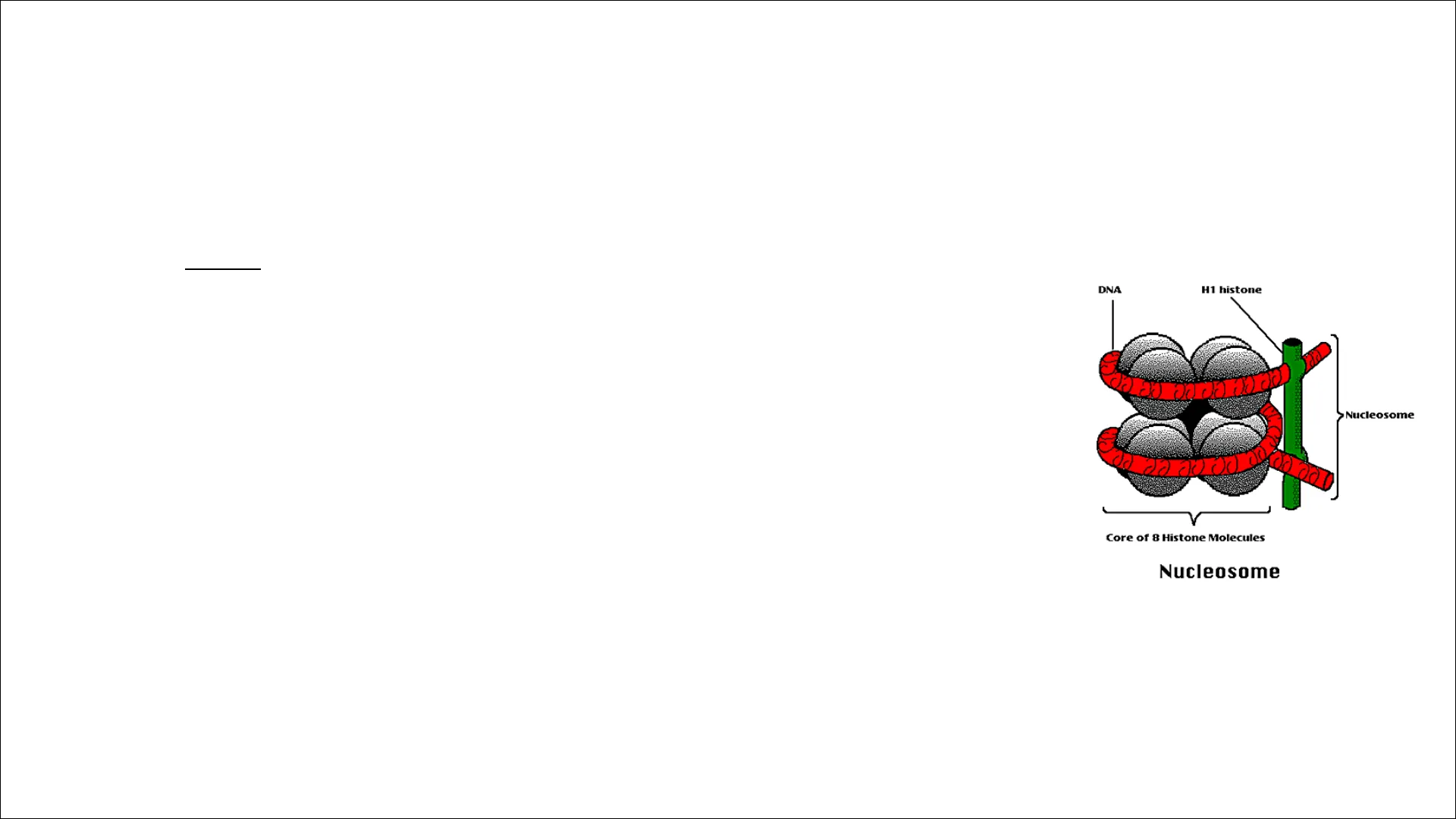
. Two copies of each four histones H2A, H2B, H3, and H4 form octamer
- Linker histone H1 basically locks the DNA in place onto the nucleosome
DNA H1 histone Nucleosome S Core of 8 Histone Molecules NucleosomeNucleosomes are the basic unit of Eukaryotic chromosome structure
- The proteins that bind to the DNA to form eukaryotic chromosomes are the histones and the non-histone chromosomal proteins,
. All four histones that make up the core of the nucleosome are relatively small proteins (102-135 amino acids) . Double-stranded DNA loops around 8 histones twice (around 140 base pairs), forming the nucleosome, which is the building block of chromatin packaging . After a short (20 to 60 base pair) "spacer" segment of DNA, the next core DNA complex forms, and so on, giving chromatin the appearance of beads on a stringNucleosomes are the basic unit of Eukaryotic chromosome structure
- In assembling a nucleosome, the histone first bind to each other to form H3-H4 and H2A-H2B dimers, and the H3-H4 dimers combine to form tetramers.
- An H3-H4 tetramer then further combines with two H2A-H2B dimers to form the compact octamer core, around which the DNA is wound.Nucleosomes are the basic unit of Eukaryotic chromosome structure
. Each of the core histones contains an N-terminal tail, which is subject to several forms of covalent modification . Histones H2A and H2b form a dimer through an interaction known as the "handshake." Histones H3 and H4 form a dimer through the same type of interaction.
- All eight N-terminal tails of the histones protrude from the disc-shaped core structure. they serve as binding sites for sets of other proteins.
- They are subject to several different types of covalent modifications that in turn control critical aspects of chromatin structure and function(A) H2A N C H2B N C H3 N C H4 N C N-terminal tail histone fold (B) (D) N N ... C N histone octamer N N N N N N (C) C C N N ...M DNA double helix side view edge view C histone H2A histone H2B histone H3 histone H4
Chromosome Structure and DNA Packaging
Chromosome Structure
- The double helix of DNA is highly negatively charged due to all the negatively charged phosphates in the backbone.
. Histones are positively charged proteins that wrap up DNA through interactions between their positive charges and the negative charges of DNA.Chromosome Structure
- DNA can be further packaged by forming coils of nucleosomes, called chromatin fibers.
- These fibers are condensed into chromosomes during mitosis, or the process of cell division.
- However, packaging of chromatin into chromosomes that we are most familiar with occurs only during a few stages of mitosis. Most of the time, DNA is loosely packaged.Chromosome Structure
Chromosome contain very tightly wound DNA You must unwind the DNA . . . and unwind the DNA . . . many times to observe the DNA double helix. 1 Histones - protein DNA wraps around.Chromosome Structure Packaging of DNA into chromosomes involves several orders of DNA coiling and folding
- Primary coiling of DNA double helix
- Secondary coiling around spherical histone "beads" forming nucleosomes
- Tertiary coiling of nucleosomes to form chromatin fibers. This allows the very long DNA molecules to fit into the cell nucleus.
- That form long loops on a scaffold of non-histone acidic proteins, that will coil tighter to form chromosome
Nucleosome Histone Charles 2 nm 11 nm 30 nm Nucleosomes Chromatine 300 nm Chromatine loops 700 nm Condensed chromatine loops 1400 nm DNA Chromosome Structure
- The interface between DNA and histone is extensive: 142 hydrogen bonds are formed between DNA and the histone core in each nucleosome.
- Nearly half of these bonds form between the amino acid backbone of the histones and the sugar-phosphate backbone of the DNA.
- Numerous hydrophobic interactions and salt linkages also hold DNA and protein together in the nucleosome.
. More than one- fifth of the amino acids in each of the core histones are either lysine or arginine (two amino acids with basic side chains)Chromosome Structure
- Nucleosome unwraps from each end at a rate of about four times per second, remaining exposed for 10 to 50 milliseconds before the partially unwrapped structure recloses.
- Eukaryotic cells contain a large variety of ATP-dependent chromatin remodeling complexes.
Hope you finished condensing your DNA, we're dividing in 10 minutes ! Awwww ...Chromosome Structure
. The nucleosome sliding catalyzed by ATP- dependent chromatin remodeling complexes. Using the energy of ATP hydrolysis, the remodeling complex is thought to push on the DNA of its bound nucleosome and loosen its attachment to the nucleosome core.
- Each cycle of ATP binding, ATP hydrolysis, and release of the ADP and Pi products thereby moves the DNA with respect to the histone octamer
--- --- ATP-dependent chromatin remodeling complex ---- ATP ADP --- CATALYSIS OF NUCLEOSOME SLIDING
Chromosome Functions
Chromosomes Functions
- Contain genetic material (DNA) which provide the genetic information for various cellular functions essential for growth, survival, development, reproduction, etc., of organisms.
- Protect the genetic material (DNA) from being damaged during cell division. Chromosomes are coated with histones and other proteins which protect it from both chemical (e.g., enzymes) and physical forces.
- Centromeres of chromosomes perform an important function in chromosome movements during cell division which is due to the contraction of spindle fibres attached to the centromeric regions of chromosomes.
- Gene expression is believed to be regulated through histone and non-histone proteins associated with chromosomes.
Genome Conservation and Gene Function
Genome conservation
- Homologous genes: Are genes similar in both their nucleotide sequence and function because of a common ancestry can often be recognized across vast phylogenetic distances
- Homologs of many human genes are present in organisms as diverse as nematode worms, fruit flies, yeasts, and even bacteria.
- Recognition of sequence similarity has become a major tool for inferring gene and protein function.
Genome conservation
- It is possible to predict the function of genes in humans for which no biochemical or genetic information is available simply by comparing their nucleotide sequences with the sequences of genes that have been characterized in other more readily studied organisms.
- Genome comparisons to search for DNA sequences that are closely similar between different species, on the principle that DNA sequences that have a function are much more likely to be conserved than those without a function
Genome conservation
- Similar pieces of DNA sequence are known as conserved regions
- These conserved regions will include regulatory DNA sequences as well as DNA sequences with functions that are not yet known. In contrast, most nonconserved regions will reflect DNA whose sequence is much less likely to be critical for function.
- The power of this method can be increased by including in such comparisons the genomes of large numbers of species whose genomes have been sequenced, such as rat, chicken, fish, dog, and chimpanzee, as well as mouse and human.
Analysis of Human Genetic Variations
Analysis of variations among human
- Errors in DNA replication, DNA recombination, or DNA repair can lead either to simple local changes in DNA sequence called point mutations such as the substitution of one base pair for another or to largescale genome rearrangements such as deletions, duplications, inversions, and translocations of DNA from one chromosome to another
- Such mutations disrupt the function or alter the regulation of existing genes.
Analysis of variations among human
- When comparing any individual human with the standard reference genome in the database, will generally find roughly 100 differences involving gain or loss of long sequence blocks
- Copy number variations (CNVs) will be very common, presumably reflecting relatively ancient origins, while others will be present in only a small minority of people
- CNVs have been implicated in many human traits, including color blindness, infertility, hypertension, and a wide variety of disease susceptibilities
- Single-nucleotide polymorphisms (SNPs) These are simply points in the genome sequence where one large fraction of the human population has one nucleotide, while another substantial fraction has another
Analysis of variations among human
- polymorphism, the variants must be common enough to give a reasonably high probability that the genomes of two randomly chosen individuals will differ at the given site; a probability of 1% is commonly chosen as the cut-off.
- SNPs in the human genome can be extremely useful for genetic mapping analyses, in which one attempts to associate specific traits (phenotypes) with specific DNA sequences (Genotype) for medical or scientific purposes
- SNPs, should therefore be rare
Analysis of variations among human
- Single nucleotide change that alters one amino acid in a protein can cause a serious disease, as for example in sickle-cell anemia, which is caused by such a mutation in hemoglobin
- A major challenge in human genetics is to recognize those relatively few variations that are functionally important against a large background of variation that is neutral and of no consequence.
- Because of purifying selection, the comparison of the genome sequences of multiple related species is an especially powerful way to find DNA sequences with important functions.
Can’t find what you’re looking for?
Explore more topics in the Algor library or create your own materials with AI.
