Anatomia dei denti umani: dentizione decidua e permanente
Slide dall'Università di Parma su Anatomia dei Denti Umani. Il Pdf esplora l'anatomia dentale, la dentizione decidua e permanente, con radiografie e diagrammi esplicativi, utile per lo studio della Biologia a livello universitario.
Mostra di più46 pagine


Visualizza gratis il Pdf completo
Registrati per accedere all’intero documento e trasformarlo con l’AI.
Anteprima
APPARATO STOMATOGNATICO
DENTAL HYGIENE : MORPHOLOGY AND FUNCTION STOMATOGNATHIC APPARATUS
ANATOMIA DEI DENTI UMANI
HUMAN THEET ANATOMY
TAS STUDI IDIORUA PARMENSIC
Prof. Paolo Vescovi DDS, PhD, MSc, Spec Oral Surgery MA · UNIVERSIT Dasser and · PARMENSIS UNIVERSITÀ DI PARMA Full Professor in Odontostomatologic Diseases Director European Master Degree in Oral Laser Applications (EMDOLA ) Director School of Speciality in Oral Surgery University of Parma
UNIVERSITÀ DI PARMA
EMDOLA European Master Degree in Oral Laser Applications
DENTI
Dentizione decidua
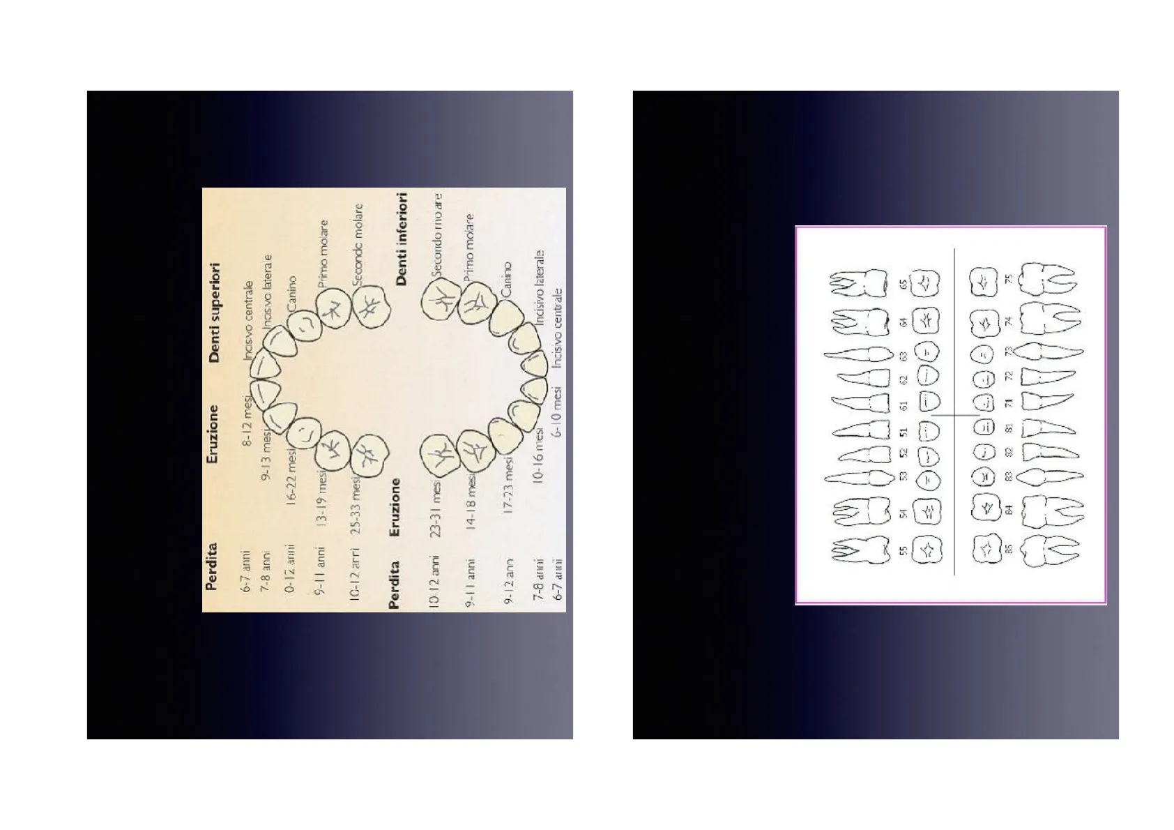
TEETH Deciduous dentition In humans there are two consecutive dentitions with two sets of teeth: deciduous (or milk) and permanent. There are 20 deciduous teeth in total, 10 per arch and 5 per quadrant. They erupt in the mouth from 6 months to approximately 2 and a half years. These teeth remain in the mouth until the age of 6. Afterwards the roots of the deciduous teeth undergo a resorption process which leads, at various times, to the loss of these teeth. The process begins around 6-7 years old with central incisors and ends around 12-13 with molars and canines. In each quadrant there are: central incisor, lateral incisor, canine and first deciduous molar, second deciduous molar.
CITAS . STUD UDIORUDeciduous dentition
Diagramma di eruzione e perdita dei denti decidui
Diagram with eruption and leak times
Perdita Eruzione Denti superiori
6-7 anni 8-12 mesi Incisivo centrale
7-8 anni 9-13 mesy Incisivo laterale
0-12 anni 16-22 mesi/ Canino
L' 9-11 anni 13-19 mesi Primo moiare
10-12 arri 25-33 mesi/ Secondo molare
Perdita Eruzione Denti inferiori
10-12 anni 23-31 mes Secondo mo are
9-1 1 anni 14-18 mesi Primo molare
9-12 ann 17-23 mesi Canino
7-8 anni 10-16 mesi Incisivo laterale
6-7 anni 6-10 mesi Incisivo centrale
Nomenclatura della dentizione decidua
Deciduous dentition - NOMENCLATURE Quadrants from 5 to 8 clockwise starting from the upper right + tooth number from 1 to 5 starting from the central incisor
C = 85 84 83 82 81 71 72 73 74 75 3 55 54 53 52 51 61 62 63 61 65 =
DENTIZIONE PERMANENTE
kPERMANENTE DENTITION There are 32 permanent teeth in total, 16 per arch, 8 per quadrant. In each quadrant there are: central incisor, lateral incisor, canine, first and second premolars (which replace the deciduous molars), first, second and third molars.
ARCATA SUPERIORE 11 21 12 22 23 :4 24 15 25 26 16 17 1 2 28 sinistra 4.8 4 3 47 37 45 36 4h 35 44 34 43 33 42 41 31 32 ARCATA INFERIORE
Nomenclatura della dentizione permanente
NOMENCLATURE: The most common system uses the combination of a number from 1 to 4 indicating the quadrant plus the tooth number from 1 to 8 starting from the central incisor.
EXERCICE 27 destra 18 38Tooth Orientation
| Centro dell'arrata dantala Vestibolare Labiale MESIAL Maciale Falutulu Palatal A Anccal A PALATAL VESTIBULAR Distale DISTAL Orale 1 Linguale Iricisale I inguala Musiale Distale Boscare LabialeO 59 7800 C5 7:00
Introduzione alla morfologia dentale
Introduction There is a certain amount of variation among individual teeth. Every tooth may not meet all the criteria for identification. By understanding the characteristics of each tooth, you will be able to differentiate among teeth, as well as between the left teeth and the right teeth in any particular group. Copyright 2003, Elsevier Science (USA). All rights reserved.
Usi clinici della morfologia dentale
Clinical Uses for Tooth Morphology Mounting dental radiographs. Assisting in charting a mouth with missing teeth and teeth that have "drifted." Selecting temporary crowns from a box with a variety of shapes. Forming matrix bands before application. Copyright 2003, Elsevier Science (USA). All rights reserved.
Dentizione anteriore permanente
Anterior Permanent Dentition 2 There are 12 anterior teeth in the permanent dentition, six in each dental arch. ? The permanent anterior teeth include the central incisors, lateral incisors, and canines. ? The central incisors are closest to the midline, the lateral incisors are the second teeth from the midline, and the canines are the third teeth from the midline. All anterior teeth are succedaneous teeth, replacing primary teeth of the same type.
Incisivo centrale Incisivo laterale Canino 1° Premolare 2° Premolare 1º Molare 2º Molare 3° Molare Copyright 2003, Elsevier Science (USA). All rights reserved.
Caratteristiche dei denti anteriori permanenti
Characteristics of Permanent Anterior Teeth 7 All anterior teeth have a cingulum, a rounded, raised area on the cervical third of the lingual surface. @ The cingulum corresponds to the lingual developmental lobe. ? The lingual surface on anterior teeth has rounded, raised borders on the mesial and distal surfaces called marginal ridges. ? Some anterior teeth have a fossa, which is a wide, shallow depression on the lingual surfaces. Copyright 2003, Elsevier Science (USA). All rights reserved.
Fig. 12-3 Various views of a newly erupted permanent maxillary incisor showing its features. Imbrication lines Cingulum Marginal ridges Mesial vincisal angle Lingual fossa DistalMamelons incisal angle Labial Incisal Lingual ridge Distal incisal angle - Mesial incisal angle Incisa Height of contour Height of contour Mesial Distal Copyright @ 2003. Elsevier Science (USA). All rights reserved. Copyright 2003, Elsevier Science (USA). All rights reserved. 2 Fig. 12-3
Incisivi centrali mascellari
Maxillary Central Incisors @ The maxillary central incisors (#8 and #9) have unique anatomic features. They are larger in all dimensions, especially mediodistally, compared with a permanent mandibular central incisor. @ The labial surfaces are more rounded from the incisal aspect, with the tooth tapering toward the lingual. The root is short compared to roots of other permanent maxillary teeth. All lingual surface features, including the marginal ridges, lingual fossa, and cingulum, are more prominent on the maxillary central incisor than on the mandibular central incisor. Copyright 2003, Elsevier Science (USA). All rights reserved.
Fig. 12-4 Various views of a maxillary right central incisor. Labial D M Lingual Incisal Mesisl Distal Copyright 2003, Elsevier Science (USA). All rights reserved. Copyright 2003, Elsevier Science (USA). All rights reserved. ? Fig. 12-4
Bordi incisali e mammelloni degli incisivi centrali mascellari
Maxillary Central Incisors- cont'd @ The incisal edges of these teeth are formed at the labioincisal line angle and do not exist until an edge has been created by wear. @ The incisal edge is also known as the incisal surface or incisal plane. @ When newly erupted, the central and lateral incisors have three mamelons, or rounded enamel extensions on the incisal ridge, or edge. The mamelons usually undergo attrition shortly after eruption. Copyright 2003, Elsevier Science (USA). All rights reserved.
Fig. 12-5 The mamelons are the rounded portions of the incisal edge of these lower central incisors. Copyright @ 2003, Elsevier Science (USA). All rights reserved. Copyright 2003, Elsevier Science (USA). All rights reserved. Fig. 12-5
Incisivi laterali mascellari
Maxillary Lateral Incisors @ The maxillary lateral incisors (#7 and #10) are smaller than the central incisors in all dimensions except root length. @ They usually erupt after the maxillary central incisors. ? The crown of a maxillary lateral incisor has a single root that is relatively smooth and straight but may curve slightly to the distal. @ Recognizing this feature is helpful when mounting radiographs. Copyright 2003, Elsevier Science (USA). All rights reserved.
Variazioni e problematiche degli incisivi laterali mascellari
Maxillary Lateral Incisors- cont'd The lateral incisors vary in form more than any other tooth in the mouth, except the third molars, and frequently are congenitally missing. ? Because of the variations in form, the permanent maxillary lateral incisors present challenges during preventive, restorative, and orthodontic procedures. 2 Often, unattractive open contacts (spaces between teeth) called diastemas occur in this area because of the variations in tooth size and position in the arch. Copyright 2003, Elsevier Science (USA). All rights reserved.
Fig. 12-6 Various views of a maxillary right lateral incisor. Labial D M Lingual Indisal Mesial Distal Copyright 8: 2003. Elevier Science (USA). All rights reserved. ? Fig. 12-6 Copyright 2003, Elsevier Science (USA). All rights reserved. Fig. 12-7 Pegged maxillary lateral incisor. Note concial shape. Copyright @ 2003, Elsevier Science (USA). All rights reserved. Copyright 2003, Elsevier Science (USA). All rights reserved. Fig. 12-7
Incisivi permanenti mandibolari
Mandibular Permanent Incisors @ The permanent mandibular incisors are the smallest teeth of the permanent dentition and the most symmetric. @ The central and lateral incisors of the mandibular arch resemble each other. ? Generally, the lateral incisor is larger than the central incisor, in contrast to the teeth in the maxillary arch. 2 Supragingival tooth deposits, such as plaque, calculus, and stain, tend to collect in the lingual concavity of the mandibular incisors. Copyright 2003, Elsevier Science (USA). All rights reserved.
Fig. 12-9 Various views of a mandibular right lateral incisor. Labial M D Lingual incisal Mesial Distal Copyright. : 2006, Elsevier Science (USA). All rights reserved. Copyright 2003, Elsevier Science (USA). All rights reserved. 2 Fig. 12-9
Incisivi centrali mandibolari
Mandibular Central Incisors @ The mandibular central incisors (#24 and #25) are the smallest and simplest teeth and are bilaterally symmetric. ? They have a small centered cingulum, subtle lingual fossa, and equally subtle marginal ridges. ? The crown of a mandibular central incisor is narrower on the lingual surface than on the labial surface. @ The developmental horizontal lines on anterior teeth, or imbrication lines, and developmental depressions are usually not present or very faint. Copyright 2003, Elsevier Science (USA). All rights reserved.
Fig. 12-8 Various views of a mandibular right central incisor. Labial M D Lingual Incisal Mesial Distal Copyright @ 2003, Elsevier Science (USA). All rights reserved. Copyright 2003, Elsevier Science (USA). All rights reserved. 2 Fig. 12-8
Incisivi laterali mandibolari
Mandibular Lateral Incisors @ The mandibular lateral incisors (#23 and #26) are slightly larger than the mandibular central incisors but otherwise are similar. @ The lateral teeth usually erupt after the mandibular central incisors. 2 The lateral incisors have a small, distally placed cingulum. @ The greater height of the cementoenamel junction (CEJ) curvature on the mesial surface than on the distal surface helps to distinguish the right mandibular lateral incisor from the left incisor. Copyright 2003, Elsevier Science (USA). All rights reserved.
Canini permanenti
Permanent Canines @ The permanent canines are the four anterior teeth located at the corners of each quadrant for each dental arch. @ Their name is derived from the Latin word for dog (canus) because these teeth resemble dogs' teeth. Patients often complain of the normal slightly deeper yellow color of their canines compared with their incisor teeth. 2 The permanent canines are the longest teeth in the dentition. Copyright 2003, Elsevier Science (USA). All rights reserved.
Non hai trovato quello che cercavi?
Esplora altri argomenti nella Algor library o crea direttamente i tuoi materiali con l’AI.
