Esercitazione pratica su organi isolati in vitro, Università di Siena
Slide dall'Università degli Studi di Siena su esercitazione pratica con organi isolati in vitro. Il Pdf, utile per corsi universitari di Biologia, esplora l'attività meccanica del muscolo liscio vascolare e i meccanismi di coupling farmaco-meccanico ed elettromeccanico.
Mostra di più47 pagine

Visualizza gratis il Pdf completo
Registrati per accedere all’intero documento e trasformarlo con l’AI.
Anteprima
Roadmap
- Attività meccanica del muscolo liscio vascolare
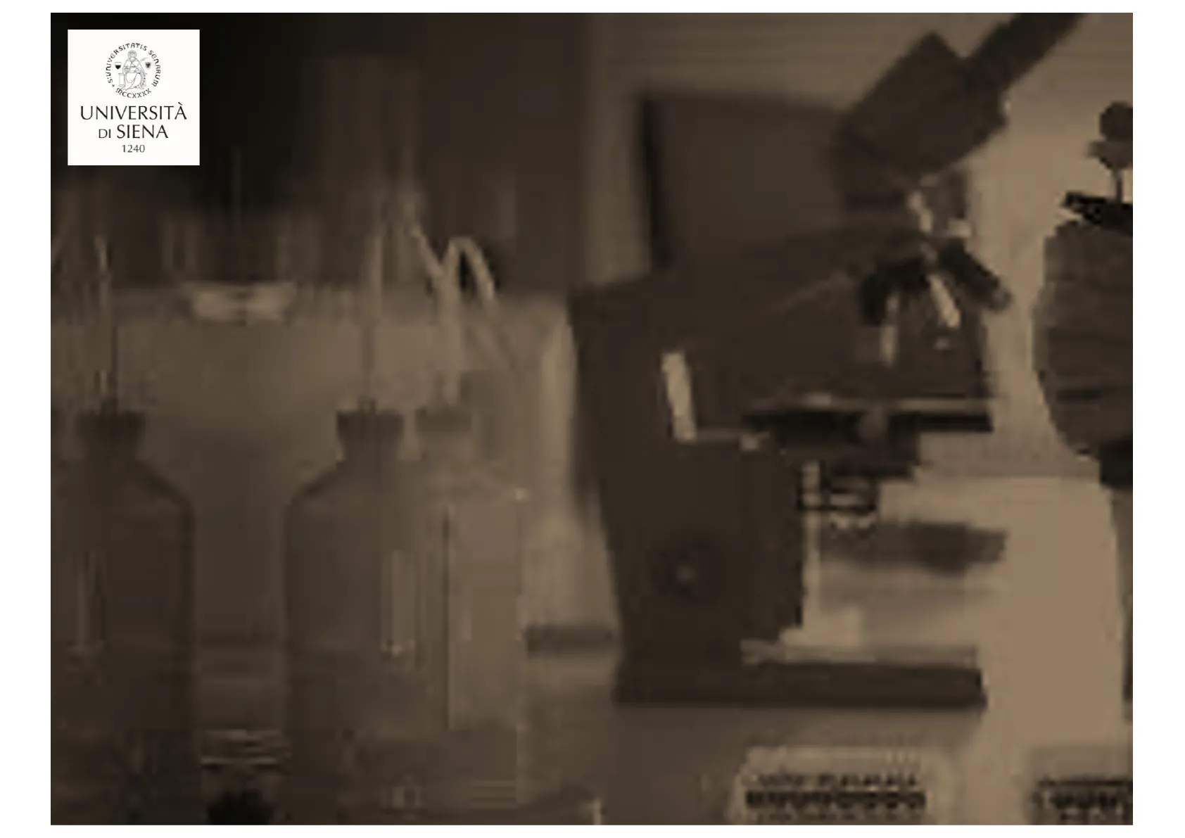
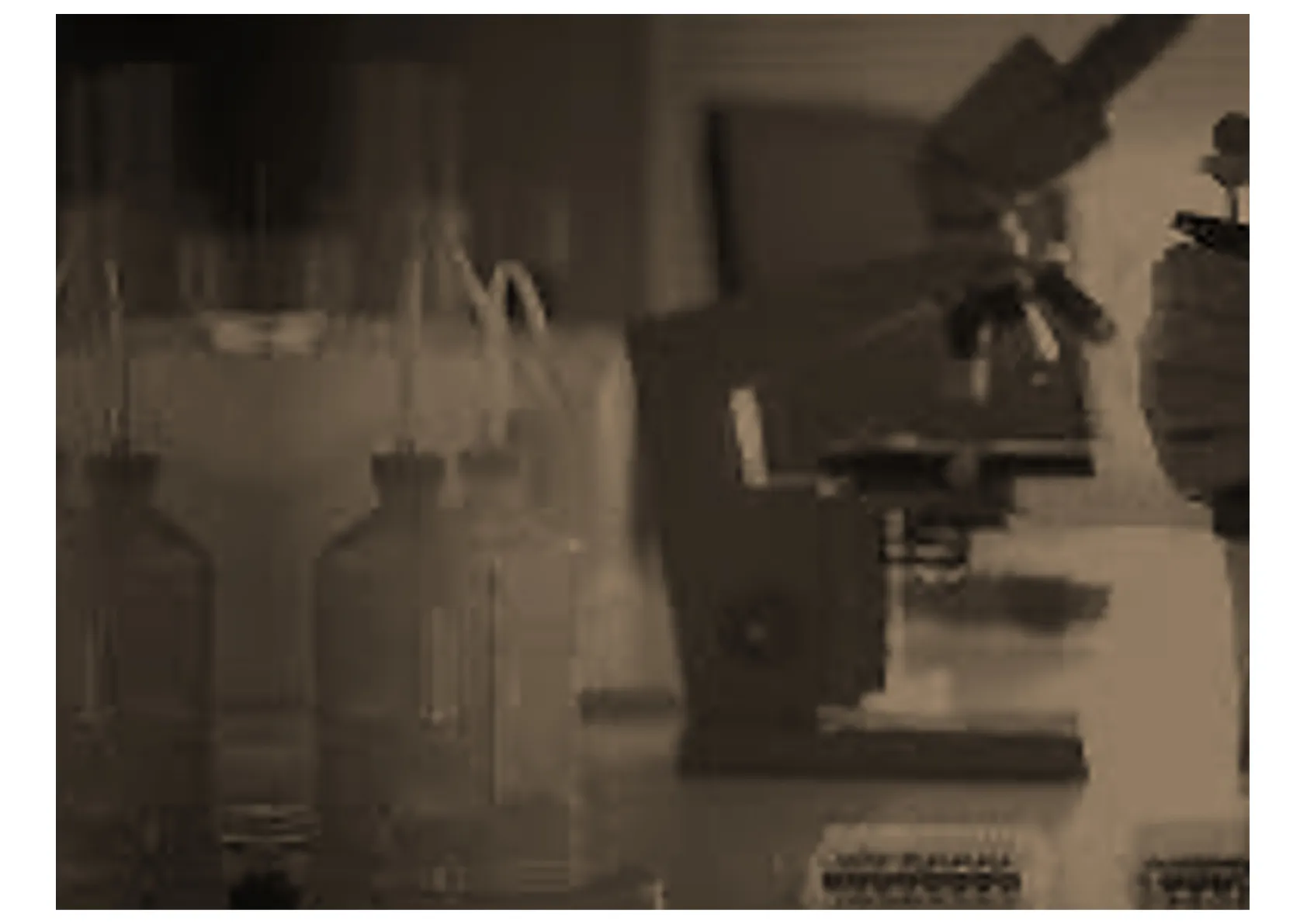
- Esercitazioni pratiche nel laboratorio di Saggi e
Dosaggi Farmacologici
Attività Meccanica del Muscolo Liscio Vascolare
Meccanismi di Contrazione e Rilassamento
- Attività meccanica del muscolo liscio vascolare
Meccanismi di contrazione e rilassamento
Il potenziale di membrana e la depolarizzazione
Via del NO - Esercitazioni pratiche nel laboratorio di Saggi e
Dosaggi Farmacologici
Meccanismo di Contrazione della Muscolatura Liscia Vascolare
Ca2+
ROC
SOC
VDCC
IP3R
+
Calmodulin
SR
2
RyR
Calmodulin
3
MLCK
Actin
ADP
ATP
4
PO4
MLC
MLCP
Fig. 3. Increased intracellular calcium stimulates contraction of vascular smooth
muscle cells 1. Mechanical or pharmacological activation increases the intracellular
calcium (Ca2+) concentration either from internal stores (sarcoplasmic reticulum, SR)
or by influx into the cell following opening of calcium channels in the plasma mem-
brane; 2. The intracellular free calcium ions bind to calmodulin and the calcium-
calmodulin complex activates myosin light chain kinase (MLCK); 3. Activated MLCK
phosphorylates the myosin light chain (MLC), which leads to cross-bridge formation
between the myosin heads and the actin filaments; 4. Cross-bridge formation results in
contraction of the smooth muscle cell. Calcium channels: Receptor-operated channel
(ROC); store-operated channel (SOC); voltage-dependent calcium channel (VDCC).
IP3R: inositol 1,4,5-trisphosphate (IP3) receptor-mediated calcium release. RyR: rya-
nodine receptor-mediated calcium release. MLCP: myosin light chain phosphatase.
Zhao et al. J Pharmacol Sci 129, 2015, 83
izzazione
Smooth muscle cell
1
Ca2+1La calmodulina
Ca2+ ions
lon channel
Calmodulin/Ca2+
100 Å
Figure 5. Ca2+ Acts Locally
Ca2+ enters cells via ion channels at rates of ~106/s, resulting in a
steep gradient of [Ca2+] (red) lasting less than 1 ms. Intracellular [Ca2+]
falls from ~10 µM to ~100 nM over a few hundred Å, a volume con-
taining >10 calmodulin molecules at normal cytosolic concentrations.
Hypothetical Ca2+ channel based on dimensions of tetrameric Kv1.2
(Long et al., 2005).
Calmodulin is a small, ubiquitous
adaptor protein that amplifies Ca2+'s diminutive size to
the scale of proteins. No other molecule more
dramatically emphasizes the evolutionary importance of
Ca2+ signaling.
Contraction of the Vascular Smooth Muscle
The Ca2+-tension relationship may change during the time course of
the contraction; the sustained phase of the contraction is maintained
by a relatively lower level of [Ca2+]i. When a greater contraction is
produced for a given elevation of [Ca2+]i, this phenomenon is referred
to as "Ca2+ sensitization of the contractile apparatus" or "an increase
in the Ca2+ sensitivity".
Contractile
stimulation
[Ca2+]¡
Ca2+ sensitivity
contraction
Fig. 1. Dual regulation of the contraction of the vascular smooth
muscle by the Ca2+ signal and the alteration of the Ca2+ sensitivity of
the contractile apparatus.
Ca2+ Sensitization
Ca2+ sensitization of the contractile proteins is signaled by the
RhoA/Rho kinase pathway
to inhibit the
dephosphorylation of the
light chain of myosin by
myosin phosphatase,
thereby maintaining force
generation.
Rho kinase inhibition
induces relaxation of
isolated segments of
smooth muscle contracted
to many different agonists.
(Webb, 2003)
agonists (norepinephrine,
angiotensin II, endothelin-1, etc.)
Ca2+
receptor
Ga
phospho-
lipase C
3 7
RhoGEF
sarcoplasmic
reticulum
+IP3
DG
Ca2+
PKC
RhoA-GTP
(active)
RhoA-GDP
(inactive)
Ca2+ -+ Ca2+/calmodulin
1
MLC kinase
(active)
Rho-
kinase
ATP
1
actin + MLC (P)
(contracted)
P
1
myosin
phosphatase
(active)
myosin
phosphatase
(inactive)
MLC
(relaxed)
FIG. 1.
Regulation of smooth muscle contraction. Various agonists (neurotransmitters, hormones, etc.) bind to
specific receptors to activate contraction in smooth muscle. Subsequent to this binding, the prototypical
response of the cell is to increase phospholipase C activity via coupling through a G protein. Phospholipase
C produces two potent second messengers from the membrane lipid phosphatidylinositol 4,5-bisphosphate:
diacylglycerol (DG) and inositol 1,4,5-trisphosphate (IP3). IP3 binds to specific receptors on the sarcoplasmic
reticulum, causing release of activator calcium (Ca2+). DG along with Ca2+ activates PKC, which phosphor-
ylates specific target proteins. In most smooth muscles, PKC has contraction-promoting effects such as
phosphorylation of Ca2+ channels or other proteins that regulate cross-bridge cycling. Activator Ca2+ binds
to calmodulin, leading to activation of myosin light chain kinase (MLC kinase). This kinase phosphorylates
the light chain of myosin, and, in conjunction with actin, cross-bridge cycling occurs, initiating shortening
of the smooth muscle cell. However, the elevation in Ca2+ concentration within the cell is transient, and the
contractile response is maintained by a Ca2+-sensitizing mechanism brought about by the inhibition of
myosin phosphatase activity by Rho kinase. This Ca2+-sensitizing mechanism is initiated at the same time that
phospholipase C is activated, and it involves the activation of the small GTP-binding protein RhoA. The
precise nature of the activation of RhoA by the G protein-coupled receptor is not entirely clear but involves
a guanine nucleotide exchange factor (RhoGEF) and migration of RhoA to the plasma membrane. Upon
activation, RhoA increases Rho kinase activity, leading to inhibition of myosin phosphatase. This promotes
the contractile state, since the light chain of myosin cannot be dephosphorylated.
voltage-
operated and
receptor-
operated Ca2+
channels
Relaxation of the Vascular Smooth Muscle
agonists
removed
channels
closed
Ca2+
receptor
2000
58
Ga
BY
phospho-
lipase C
Ca,Mg-
ATPase
Na+/Ca2+
exchanger
+ Ca2+
binding proteins
+
Ca2+
Ca2+/calmodulin
Ca,Mg-ATPase
MLC kinase
(active)
1
Ca2+
actin + MLC (P)
(contracted)
MLC
phosphatase
(active)
MLC
(relaxed)
FIG. 2.
Relaxation of smooth muscle. Smooth muscle relaxation occurs either as a result of removal of the
contractile stimulus or by the direct action of a substance that stimulates inhibition of the contractile
mechanism. Regardless, the process of relaxation requires a decreased intracellular Ca2+ concentration and
increased MLC phosphatase activity. The sarcoplasmic reticulum and the plasma membrane contain Ca,Mg-
ATPases that remove Ca2+ from the cytosol. Na+/Ca2+ exchangers are also located on the plasma membrane
and aid in decreasing intracellular Ca2+. During relaxation, receptor- and voltage-operated Ca2+ channels in
the plasma membrane close resulting in a reduced Ca2+ entry into the cell.
Ca2+
voltage-
operated and
receptor-
operated Ca2+
channels
sarcoplasmic
reticulum
Meccanismi Principali di Controllo della Contrazione Muscolare Liscia
Nifedipina
CONTRAZIONE
RILASCIAMENTO
Agonisti
Noradrenalina
Istamina
Angiotensina
ecc.
Bloccanti dei
canali del calcio
Attivatori dei canali
del potassio
(cromakalim ecc.)
Inibitori
PDE
Agonisti
Adenosina
ß-agonisti
Prostaglandine
ecc.
ATP
+
Ca2+ Ca2+ Na+
ANP
NO
I
1
2
3
4
5
6
PLC
AC
GC
+
+
+
+
+
7
IP3
8
PDE
CAMP
CGMP
+
+
DEPOLARIZZAZIONE
IPERPOLARIZZAZIONE
PKA
PKG
Rilascio di Ca2+
İ[Ca2+]
CONTRAZIONE
CELLULA MUSCOLARE LISCIA
O
+
GC
1
Misurazione del Potenziale di Membrana
K+mV
0
0
A
-100-
100
millivolt
-100-
voltmetro
elettrometrico
t
microelettrodo
elettrodo di
riferimento
+
1
-
-
+
+
-
-
+cellula
+
mV
0
0
-
-100
-100
millivolt
-100
1
t
microelettrodo
elettrodo di
riferimento
+
+
1
+
-
-
+
-
-
+cellula
+
Misurazione del
potenziale di
membrana
All'elettrodo di riferimento
viene assegnato un valore di
0 mV
B
voltmetro
elettrometrico
Generation of Resting Membrane Potential
IGeneration of resting membrane potential (Wright, 2004)
How does the electrical gradient arise?
1.the presence of large gradients for K+ (outwardly directed) and Na+
(inwardly directed) across the plasma membrane (the product of the
activity of the Na+/K+-ATPase)
2.the relative permeability of the membrane to those ions (the open vs.
closed status of ion-selective membrane channels).
inside
Outside
100 mM KCl
10 mM KCl
10 mM NaCl
100 mM NaCl
Membrane permeable only to K+Chemical force
inside
Outside
100 mM KCl
10 mM KCl
10 mM NaCl
100 mM NaCl
Cl-
K+
Cl- K+
Cl- K+
Membrane permeable only to K+Electrical force
inside
Outside
100 mM KCl
10 MM KCI
10 mM NaCl
100 mM NaCl
K+
Cl-
Cl- K+
Cl-
K+
K
Membrane permeable only to K+Chemical force = Electrical force
inside
Outside
100 mM KCl
10 mM KCl
10 mM NaCl
100 mM NaCl
Cl-
Cl- K+
Cl-
K+
K+
本 本 本
Membrane permeable only to K+
Nernst Equation
Nernst equation:
VK = -
RT
zF
In
K]
in
K
outNernst equation:
VK = -
RT
zF
In
[K]in
[K]
out
where VK is the equilibrium electrical PD, which exactly
opposes the chemical energy of the chemical gradient, the
intracellular-to-extracellular K+ concentration ratio ([K];/
[K]out). R is the gas constant with units of 8.31 J/(Kmol), T is
absolute temperature in Kelvin (37°C = 310 K), F is Faraday's
constant at 96,500 coulombs/mol, and z is the valance of the
ion question; + 1 for K+. It is instructive to insert the relevant
values for R, T, F, and z, and to convert from the natural log to
the common (base 10) log by multiplying by 2.303. The Nernst
equation then becomes (at 37℃)
VK =
- 0.0615 Volts
Z
log 10[
[K]in
K]
lout
It is convenient to simplify this equation to an adequate (and
useful) approximation
VK = - 60 mV log10
[K]
[K]in
lin
lout
When we consider the K+ gradient of our example (100 mM
inside, 10 mM outside) we find that this outwardly directed
10-fold gradient of a monovalent cation is balanced by a 60 mV
electrical PD (in this case, inside negative).
PD: potential
difference
"Reversal potential"
at -70 mV the
direction of net K+
flux would reverse!
Efflux of K+ from the Cell
"If we started with 100 mM K+ inside and there was a net
efflux of K+ from the cell, shouldn't the intracellular K+
concentration now be lower?“
inside
Outside
100 mM KCl
10 mM KCl
10 mM NaCl
100 mM NaCl
Cl-
Cl- K+
K+
K+
Cl-
Membrane permeable only to K+
The amount of K+ that leaves the cell to produce the
equilibrium potential is sufficiently small that it cannot be
measured chemically, despite the substantial electrical effect
it has.
Non hai trovato quello che cercavi?
Esplora altri argomenti nella Algor library o crea direttamente i tuoi materiali con l’AI.
