Vascular Pathology: Overview of Diseases and Tumors by Prof. Maurilio Ponzoni
Document from Prof. Maurilio Ponzoni about Vascular Pathology. The Pdf explores vascular diseases, normal vessel structure, congenital anomalies, and hypertensive vascular disease. This University-level Biology document, authored by Prof. Maurilio Ponzoni, also details vascular tumors like hemangiomas and glomus tumors.
See more17 Pages

Unlock the full PDF for free
Sign up to get full access to the document and start transforming it with AI.
Preview
Vascular Pathology Overview
Nilayda Gultekin Prof. Maurilio Ponzoni Pathologic Anatomy, MP3 18/10/2023 Vascular Pathology The lecture was actually held by professor Sanvito, who will be focusing on cardiovascular pathology. This first lecture deals with vascular pathology, while the next lecture will deal with cardiac pathology. We must study on our own the pathology of veins and lymphatic vessels.
Overview of Vascular Disorders
Two principal mechanisms underlie all vascular disorders. The first one is the narrowing (stenosis) or complete obstruction of vessel lumens that can happen suddenly (thrombosis, embolism) or progressively, as in the case of atherosclerosis. The other mechanism is the weakening of the vessel walls that may lead to dilation, aneurysm or rupture of the wall. Slides: Vascular diseases include congenital anomalies, hypertensive vascular disease, atherosclerosis, aneurysms, dissection, vasculitis, tumors and many more.
Normal Vessel Structure and Function
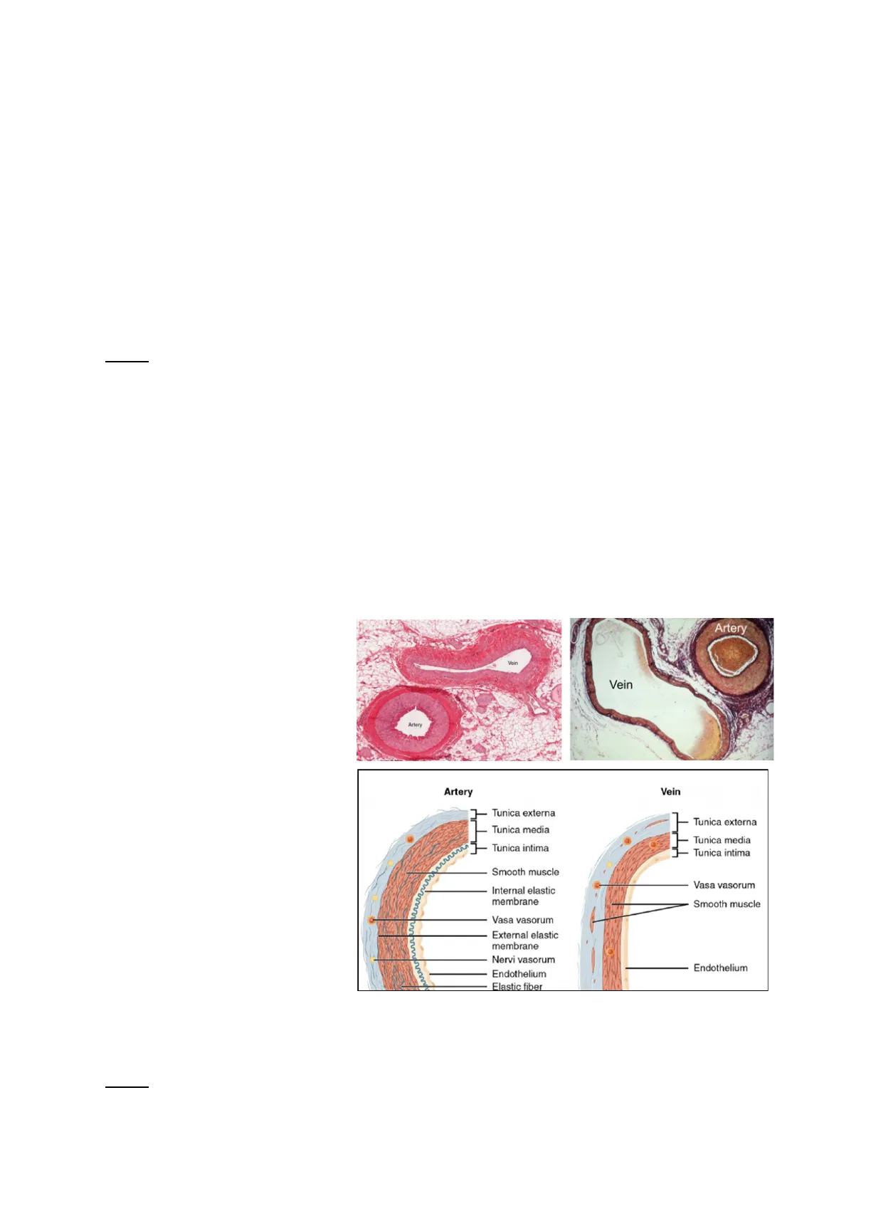
The pathogenesis of vascular diseases is better appreciated by understanding the normal structure and function of vessels. The thickness and composition of the wall depend on hemodynamic forces and body/tissue requirements. Arteries can be elastic or muscular. Large vessels - including the aorta and its branches (e.g. subclavian, carotid, iliac arteries) - are elastic arteries and have the function to accommodate the pulsatile flow and high blood pressure. Elastic arteries are able to recoil during diastole and then transmit the energy into forward blood flow during systole. Elastic arteries' tunica media are composed of lamellar units made of elastic fibers, separated by smooth muscle cells and embedded in the ECM. Medium-sized muscular arteries comprise small branches of the aorta (e.g. coronary and renal arteries) and are shown in these pictures. The left image is an H&E staining that is used to make diagnoses, while the right image is a special staining that highlights the elastic fibers. The internal elastic lamina and the external elastic lamina are defined better in arteries than in veins. Muscular arteries' media are composed of smooth muscle cells; elastic fibers are present in internal and external elastic laminae, embedded in the ECM. Artery Vein Vein Arter Artery Vein Tunica externa Tunica externa Tunica media Tunica media Tunica intima Tunica intima Smooth muscle Vasa vasorum Internal elastic membrane Smooth muscle Vasa vasorum External elastic membrane Nervi vasorum Endothelium Endothelium Elastic fiber Muscular arteries can contract and relax, influencing the blood flow. The contraction and relaxation depend on a lot of factors, being regulated by the autonomic nervous system, local factors and hormones. Slides: Small arteries (<2 mm in diameter) and arterioles (20-100 um in diameter) are present within tissues and organs.
1Nilayda Gultekin Prof. Maurilio Ponzoni Pathologic Anatomy, MP3 18/10/2023
Congenital Anomalies of Vessels
Congenital anomalies are variants of the normal anatomic pattern of vascular supply and are important during surgery, as the surgeon should know the unexpected location of the injured vessel. This is very significant for coronary arteries. Two important congenital anomalies are:
- Development aneurysms are aneurysms that can be diagnosed when they rupture and induce a hemorrhage, otherwise they are silent. (Slides: in cerebral vessels, the rupture causes fatal intracerebral hemorrhage).
- Arteriovenous fistulas are abnormal, small, direct connections between an artery and a vein, bypassing capillaries, causing a shunt of arterial and venous blood. The additional blood volume can be a problem for the heart to pump.
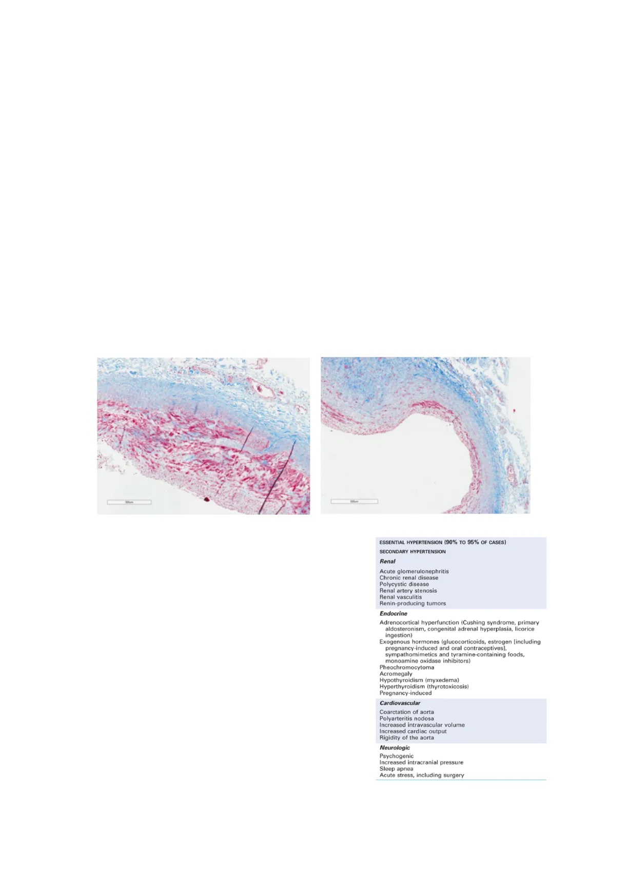
Among congenital anomalies, fibromuscular dysplasia is not very easily diagnosed and of unknown etiology, due to hormonal and mechanical factors. The media is characterized by a peculiar microscopic pattern. In the special staining below, you can see smooth muscle cells and the extracellular matrix. The media is deranged by deposition of a fibrous ECM and the smooth muscle cells are also disorganized. This constitutes a problem as the defect is often localized at renal, carotid, splanchnic or vertebral level: the thickening and stenosis of the lumen can impair the organ oxygen supply causing ischemia, infarct or hemorrhages. In later stages, we can also have dilation and aneurysm due to weakness of the wall.
Hypertensive Vascular Disease
One of the most common causes of vascular disorder is hypertension. If the diastolic pressure is 80 or more and if the systolic pressure is 140 or more, we consider it high *. Usually, cardiovascular diseases affect mostly elderly people. Only in the case of genetic diseases, they are multifactorial and affect young people. Low pressure (hypotension) may result in impaired or inadequate perfusion of the organs and tissue leading to ischemia, necrosis and tissue death. High pressure (hypertension) can cause damage to the vessel walls with dilation or rupture and end-organ damage. The table on the right shows the types and causes of hypertension. 90-95% of the cases are essential or idiopathic: the cause of hypertension is still unknown, but it is well controlled and compatible with long life. ESSENTIAL HYPERTENSION (90% TO 95% OF CASES) SECONDARY HYPERTENSION Renal Acute glomerulonephritis Chronic renal disease Polycystic disease Renal artery stenosis Renal vasculitis Renin-producing tumors Endocrine Adrenocortical hyperfunction (Cushing syndrome, primary aldosteronism, congenital adrenal hyperplasia, licorice ingestion) Exogenous hormones (glucocorticoids, estrogen [including pregnancy-induced and oral contraceptives], sympathomimetics and tyramine-containing foods, monoamine oxidase inhibitors) Pheochromocytoma Acromegaly Hypothyroidism (myxedema) Hyperthyroidism (thyrotoxicosis) Pregnancy-induced Cardiovascular Coarctation of aorta Polyarteritis nodosa Increased intravascular volume Increased cardiac output Rigidity of the aorta Neurologic Psychogenic Increased intracranial pressure Sleep apnea Acute stress, including surgery Secondary hypertension is due to renal, endocrine, cardiovascular or neurological factors.
2Nilayda Gultekin Prof. Maurilio Ponzoni Pathologic Anatomy, MP3 18/10/2023 Slides: besides increasing the risk of atherosclerosis, hypertension can cause cardiac hypertrophy, heart failure (hypertensive heart disease), multi-infarct dementia, aortic dissection and renal failure. *Prof. Ponzoni said that the physiological values of blood pressure should be less than 120 mmHg for systolic pressure and 80 mmHg for diastolic pressure. Prof. Sanvito meant that cardiologists define sustained diastolic pressure when over 89 mmHg and sustained systolic pressure when above 139 mmHg. In other words, hypertension is diagnosed when the blood pressure is consistently 140/90 mm Hg or higher. The cartoon aims to show how blood pressure is regulated. The arterial blood pressure depends on cardiac output and peripheral resistance. A lot of factors influence either the resistance or the cardiac output, such as the autonomic nervous system, autocrine hormone factors, functionality of the kidney, vessel structure and function, blood volume and myocardial contractility. Arterial pressure Total peripheral resistance x Cardiac output · Extracellular fluid volume · Blood volume · Arterial and venous compliance · Resistance to venous return Kidney structure and function Neuroendocrine Structure and function of blood vessels and heart · Pressure-natriuresis (Sympathetic nervous system, · Local autoregulation · Tubular Na* reabsorption hormones, paracrine and autocrine factors) · Vascular reactivity · Glomerular filtration rate: urinary protein . Aorta and heart-wall · Renal blood flow thickness and morphology · Renal vascular resistance · Microvessel density · Morphology T Copyright 2006 Nature Publishing Group Nature Reviews | Genetics Hypertension accelerates atherogenesis and can cause degenerative changes in the vessel wall, which can lead to aortic dissection and cerebrovascular hemorrhages.
Arteriosclerosis
Arteriosclerosis is the hardening of the arteries (from Greek) and is an aspecific, generic term reflecting the stiffness achieved by damage and loss of plasticity of vessels. There are different patterns of arteriosclerosis:
- Arteriolosclerosis affects small arteries and arterioles and causes ischemic injury to the downstream tissue. It includes hyaline and hyperplastic arteriolosclerosis.
- Atherosclerosis is the formation of cholesterol plaques in or on the arterial walls.
Hyaline Arteriolosclerosis
Kivaline arteriolosclerosis Hyaline arteriosclerosis is a homogeneous, hyaline thickening of the vessel wall withduminal narrowing Hyaline means pink. In the picture, we can see the pink small artery, the nuclei of endothelial cells, particularly thin intima layers and a homogeneous media. There is the leakage of plasma proteins across the mjured intima layer and anIncrease in smooth muscle cells. Stenosis decreases the blood flow and pressure. A Slides: the activation of the renin-angiotensin system increases peripheral resistance and blood volume through vasoconstriction direct action on SMC and aldosterone secretion with increased sodium reabsorption in the distal tubule. stenosis > narrowing of a verrel 3 · e.g. renin-angiotensin-aldosterone system, adrenaline, noradrenaline. arginine vasopressin, atrial natriuretic peptide reactive oxygen species, nitric oxide . Cardiac contractilityNilayda Gultekin Prof. Maurilio Ponzoni Pathologic Anatomy, MP3 18/10/2023
Hyperplastic Arteriolosclerosis
Hyperplastic arteriolosclerosis occurs in severe or malignant hypertension and is characterized by vessels exhibiting "onion-skin" concentric lésions, lamination of the media (wall thickening) and luminal narrowing. The consequences are ischemja and tissue necrosis. Slides: Laminations are SMCs with thickened basement membranes.
Atherosclerosis
Atherosclerosis is one of the most important vascular disorders and its risk factors include:
- Age: it occurs especially between 35 and 55 years of age.
- Sex: males are more predisposed than females.
- Genetics: there is a familial predisposition. Since there are a lot of genes involved, atherosclerosis is considered a polygenic disease.
- High levels of LDL cholesterol cause hyperlipidemia.
- Smoking.
- Diabetes.
- Elevated plasma homocysteine.
The key feature of atherosclerosis is the atheromas, atheromatous or atherosclerotic plaques. Atheromatous plaques appear as raised lesions in the lumen of vessels, usually soft, grumous and yellow at the beginning, and are characterized by a core of lipids (mainly cholesterol or cholesterol esters) with inflammatory cells and macrophages inside and covered by a white fibrous cap. In a microscopic picture of a plaque, you can see the fibrous cap and the lipid core around. The lipid is mostly found inside the macrophages and appears clear because, during the tissue preparation, we use ethanol dehydration and all lipids disappear. Slides: fatty streaks are the earliest lesions in case of atherosclerosis and are composed by lipid-filled foamy macrophages. FIBROUS CAP (smooth muscle cells, macrophages, foam cells, lymphocytes, collagen, elastin, proteoglycans, neovascularization) NECROTIC CENTER (cell debris, cholesterol crystals, foam cells, calcium) MEDIA Below, you can find two gross pictures of the aorta. The left image shows a thoracic aorta (A), where the dots represent the intercostal arteries. Yellow is not the physiological color of the aorta, but rather is given by the lipid accumulation with the formation of whitish or yellow atherosclerotic plaques. The right image shows an abnormal thoracic aorta. The brown plaques are ulcerated plaques, complicated with superimposed thrombosis.
4
Can’t find what you’re looking for?
Explore more topics in the Algor library or create your own materials with AI.
