Detection of Specific DNA Fragments Within a Genomic Sample
Slides about Detection of Specific DNA Fragments Within a Genomic Sample. The Pdf, a university-level Biology presentation, details DNA fragment detection techniques like Southern blotting and PCR, including its phases and the Real-time PCR method.
See more58 Pages


Unlock the full PDF for free
Sign up to get full access to the document and start transforming it with AI.
Preview
Detection of Specific DNA Fragments
- Southern blotting
- RFLP
- Polymerase Chain Reaction (PCR)
Southern (Edwin) Blotting Technique
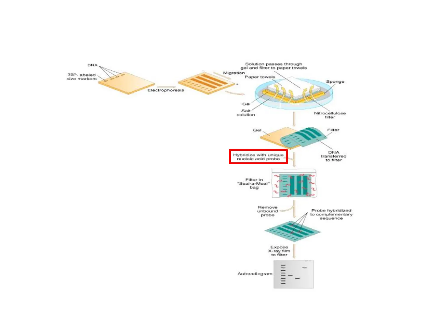
A Southern blot is a technique for transferring a DNA electropherogram from a gel onto a carrier solid phase (i.e. nitrocellulose paper). The purpose is to have DNA available for hybridization with a nucleic acid probe, detecting a given DNA sequence of interest.
Solution passes through gel and filter to paper towels
Migration Paper towels Sponge Electrophoresis Gel Salt solution Nitrocellulose filter Gol Filter Hybridize with unique nucleic acid probe DNA transferred to filter ? 2 Filter in "Seal-a-Meal" bag Remove unbound probe Probe hybridized to complementary sequence Expose X-ray film to filter - Autoradiogram - DNA 32P-labeled size markers
Southern (Edwin) Blotting Steps
- Extract and purify DNA from cells
- DNA is restricted with enzymes
- Separated by electrophoresis
- Denature DNA -
- Transfer to nitrocellulose paper (Blotting)
- Add labeled probe for hybridization to take place
- Wash off unbound probe
- Autoradiograph
1 Capillary force Weight Paper towels Membrane Gel + Buffer Electrophoresis of restriction digested DNA fragments Denaturation of DNA (~0.5 M NaOH) and blotting Sponge Visualization Washing off unbound probe Labelled marker Hybridization with labelled probe Hybridized fragments
Oligonucleotide DNA Probes
Oligonucleotide probes are short stretches of single-stranded DNA (or RNA) used to detect the presence of complementary nucleic acid sequences (target sequences) by hybridization. They are usually labelled, for example with radioisotopes or fluorophores, to enable their detection.
When a certain sequence needs to be detected in a DNA sample, a complementary oligonucleotide is synthesized in the laboratory.
Oligonucleotide probe 5' gatcatttaacgtagc3' 3' ... catcgatagctaatcgtgctagtaaattgcatcgaatcgtagctagtc ... 5' DNA sample
Design of Oligonucleotide DNA Probes: Specificity
Restriction Enzyme Cutting
3' ... catcgatagctaatcgtgctagtaaattgcatcgaatcgtagctagtc ... 5' Let's consider a DNA sample that can be cut by a restriction enzyme at the 5' t|tagca3' restriction site ... 3'aatcgt ... resulting in 3 fragments: A. 3' ... catcgatagctaatcg5' B. 3'tgctagtaaattgcatcgaatcg5' C. 3' tagctagtc ... 5'
Electrophoresis of Fragments
When submitted to electrophoresis these fragments will run as C (faster), A, B (slower).
Design of Oligonucleotide Probes: Electrophoresis
Dna fragments: A. 3' ... catcgatagctaatcg5' B. 3'tgctagtaaattgcatcgaatcg5' C. 3'tagctagtc ... 5' submitted to electrophoresis will run as C (faster), A, B (slower).
DNA size markers 1 LOAD DNA ONTO GEL AND APPLY VOLTAGE I negative electrode O - B - A - C positive electrode slab of agarose gel
NB: if you don't stain the gel with a DNA-binding dye to have the DNA fragments evidenced as a bands, representing groups of same-sized DNA fragments, you do not see any signal.
Design of Oligonucleotide Probes: Potential Probes
3' ... catcgatagctaatcgtgctagtaaattgcatcgaatcgtagctagtc ... 5' 3' ... catcgatagctaatcgtgctagtagcttgcatcgaatcgtagctagtc ... 5' 3' ... catcgatagctaatcgtgctagtagcttgcatcgaatcgtagctagtc ... 5' Potential probes: 5' atgc3' 5' gtagct3' 5' gatcatttaacgtagc3'
Design of Oligonucleotide Probes: Specificity for Southern Blotting
5' gatcatttaacgtagc3' 3' ... catcgatagctaatcgtgctagtaaattgcatcgaatcgtagctagtc ... 5' 5' atcg3' 5' atcg3' 5' atcg3' 3' ... catcgatagctaatcgtgctagtagcttgcatcgaatcgtagctagtc ... 5' 5' gtagct3' 5' gtagct3' 3' ... catcgatagctaatcgtgctagtagcttgcatcgaatcgtagctagtc ... 5' Potential probes: 5' atgc3' 5' gtagct3' 5' gatcatttaacgtagc3' good specificity Southern blotting probe
A Good, Specific, Southern Blotting Probe
DNA size markers LOAD DNA ONTO GEL AND APPLY VOLTAGE negative electrode O - positive electrode slab of agarose gel 32P. autoradiography
Restriction Fragment Length Polymorphism (RFLP)
A mirror of the use of several restriction endonuclease type when sequencing a genome is using only a specific one to cleave several genomic DNA samples. In this case a Restriction Fragment Length Polymorphism (RFLP) will likely appear, which is a difference in individual DNA sequences that can be detected by the presence of fragments of different lengths after digestion of the DNA samples.
Chromosomal DNA (e.g., Suspect 1) Cleave with restriction DNA fragments Separate fragments by agarose gel electrophoresis (unlabeled). The RFLP probes are frequently used in genome mapping such as forensics ... DNA markers DNA markers Suspect 1 Evidence Victim Suspect 2 DNA markers DNA markers Radiolabeled DNA probe I I S E V 5 E I I I I 1 I - Denature DNA, and transfer to nylon membrane. Incubate with probe, then wash. - - Expose x-ray film to membrane. - E Evidence - 1
RFLP Probe Characteristics
An RFLP probe is a labeled DNA sequence that hybridizes with a limited number of fragments of the digested DNA sample after they were separated by gel electrophoresis, thus revealing a unique blotting pattern characteristic to a specific genotype.
RFLP for Paternity Testing
RFLP (Restriction Fragment Length Polymorphism = (see Southern blotting). This genetic analysis, that allows to identify unique patterns of restriction enzyme DNA cutting is also a standard protocol to perform a paternity test.
ABCD ] ] ] - 87654321 A - Parent 1 B - Parent 2 C - Son 1 D - Son 2
Polymerase Chain Reaction (PCR) Basis
The knowledge (in a pill ... ) of how a DNA molecule naturally duplicate lies at the basis of a Polymerase Chain Reaction (PCR).
Daughter strands Origins of replication for three different replicons Parent strands Replicaton forks Replication bubbles Two replicons on left fuse together Replicons fuse
Replication Bubble
A replication bubble is an unwound and open region of DNA where DNA replication occurs. Bubbles are created when the enzyme helicase separates the two strands of DNA so that they can be replicated.
Polymerase Chain Reaction (PCR) Overview
Polymerase chain reaction (PCR) is a technique used to "amplify" segments of DNA
Dancing NAKED IN the MIND FIELD WINNER OF THE NOBEL PRIZE IN CHEMISTRY KARY MULLIS "Kary Mulis, perhaps the weirdest human ever to win the Nobel Prize in Chemistry, [has written] a chatty, rambling, funny, iconoclastic tour through the wonderland that is [tis] mind" -THE WASHINGTON POST
PCR Technique and Significance
The polymerase chain reaction (PCR) is a fast and inexpensive technique used to "amplify" - i.e. to copy - small segments of DNA. Because significant amounts of a sample of DNA are necessary for molecular and genetic analyses, studies of isolated pieces of DNA are made possible with PCR amplification. Often heralded as one of the most important scientific advances in molecular biology, PCR revolutionized the study of DNA to such an extent that its creator, Kary B. Mullis, was awarded the Nobel Prize for Chemistry in 1993.
The Nobel Prize in Chemistry 1993
"for contributions to the developments of methods within DNA-based chemistry"
Kary Mullis's Contribution
"for his invention of the polymerase chain reaction (PCR) method" Kary Mullis 1/2 of the prize USA La Jolla, CA, USA
Michael Smith's Contribution
"for his fundamental contributions to the establishment of oligonucleotide-based, site-directed mutagenesis and its development for protein studies" Michael Smith 1/2 of the prize Canada University of British Columbia Vancouver, Canada
DNA Features: Thermal Denaturation
When DNA is heated, hydrogen bonds are destabilized: the double filament separates. This step is defined thermal denaturation. Heat affects AT more then CG base pairs, since AT pairs bind more weakly (2 bonds) than GC (3 bonds) pairs.
Melting Temperature (Tm)
Melting temperature (Tm) by definition is the temperature at which one half of the DNA duplex will dissociate to become single stranded and indicates the duplex stability. Cooling allows new bonds to form between the sequences (annealing, rehybridization, renaturation)
Renaturation Step 2 (fast) Denaturation Denaturation Renaturation Step 1 (slow) Random association Dissociation Open coil
DNA Melting and Annealing Profile
100 Melting % Single-Strand DNA 80 60 40 Annealing 20 T. TM 30 40 50 60 70 80 90 Temperature ("℃)
Profile Characteristics
Melting (solid line) and annealing (broken line) profile for double-stranded DNA. The melting is a cooperative reaction. Once begun, relatively small increases in melting temperature (TM = . ) result in the rupture of relatively large number of hydrogen bonds. The melting profile differs greatly from the annealing (cooling) profile (TA = · ). The evident hysteresis in the process arises from differences in the rates of hydrogen bond breaking and specifical annealing of the complementary strands.
PCR Amplification Process
To amplify a segment of DNA using PCR, the sample is first heated so the DNA denatures, or separates into two pieces of single-stranded DNA. Next, an enzyme called "Taq polymerase" synthesizes - builds - two new strands of DNA, using the original strands as templates. This process results in the duplication of the original DNA, with each of the new molecules containing one old and one new strand of DNA.
Region to be amplified 5' 3' Target DNA 3 5 Add excess primers 1 and 2, dNTPs, and Taq polymerase Heat to 95° to melt strands Primer 1 Cool to 60° to anneal primers Cycle 1 5' 3 - Primer 2 3' 5' Primers extended by Tag polymerase at 60° 5° 3 3' 5 Heat to 95° to melt strands Cool to 60° to anneal primers 3'S 5' 5' 3' Primers extended by Tag polymerase at 60° Cycle 2 3' 5' 5' 3' Heat to 95° to melt strands Cool to 60° to anneal primers
PCR Cycling and Automation
Then each of these strands can be used to create two new copies, and so on, and so on. The cycle of denaturing and synthesizing new DNA is repeated as many as 30 or 40 times, leading to more than one billion exact copies of the original DNA segment.
Cycle 3 Primers extended by Tag polymerase at 60° Heat to 95° to melt strands Cool to 60° to anneal primers Primers extended by Taq polymerase at 60° Cycle 4 1 And so on
separate DNA strands and anneal primer DNA synthesis separate DNA strands and add primer DNA synthesis DNA oligonucleotide primers region of double-stranded chromosomal DNA to be amplified + FIRST CYCLE SECOND CYCLE 1 2 4 separate DNA etrands and anneal primer DNA synthesis etc. 1 THIRD CYCLE 8
Automated PCR with Thermocycler
The entire cycling process of PCR is automated and can be completed in just a few hours. It is directed by a machine called a thermocycler, which is programmed to alter the temperature of the reaction every few minutes to allow DNA denaturing and synthesis.
FOTO Biometra" MENS 1071 900m GRADIENT 4 1 3
Can’t find what you’re looking for?
Explore more topics in the Algor library or create your own materials with AI.
