Pathology and Diagnostics: Classical Serum Proteins and Diagnostics
Slides about Pathology and Diagnostics: Classical Serum Proteins, diagnostics. The Pdf, a presentation for university students in Biology, describes serum protein electrophoresis, focusing on albumin, alpha, beta, and gamma globulins, and their diagnostic implications.
See more22 Pages

Unlock the full PDF for free
Sign up to get full access to the document and start transforming it with AI.
Preview
Classical Serum Proteins and Diagnostics
Da Rin - Clinical Biochemistry- Lesson 08-10: Classical Serum Proteins, diagnostics 29/11/2024 - Group 45 (Martínez and Camponogara)
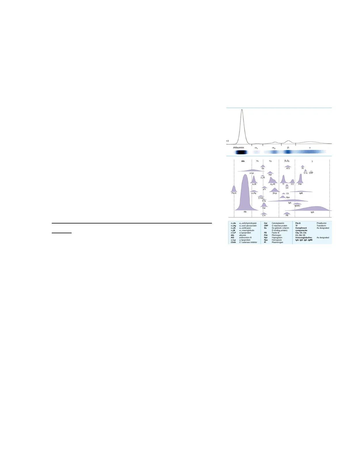
When separating proteins in the serum, we know there are specific regions where the serum proteins can migrate. In particular, we have the region of albumin, alpha-1, followed by the alpha-2, beta, and gamma regions. The beta region is further divided into beta-1 and beta-2. Within this distribution, we can identify certain proteins that make up the majority of each region.
This plot simply represents the distribution of serum proteins during electrophoresis, and it shows how to interpret the data from the separation.
The separated serum proteins typically form five main bands:
- @1-antichymotrypsin
- Cer Ceruloplasmin
- Pre A Prealbumin
- C& Ag 0.1-acid glycoprotein
- CRP C-reactive protein
- Tf Transferrin
- CL.At o41-antitrypsin
- Go Gc-globulin (vitamin Compliment As designated C1-macroglobulin D-binding protein) components:
- C-LP c-lypoprotein
- FB Factor B
- Clq, Clr, Cls
- Alb albumin
- Fibr Fibrinogen
- C3, C4, C5
- At3 antihrombin III
- Hpt Haptoglobin
- Immunoglobulins: As designated
- C-Lp at-lipoprotein
- Hpx Hemopexin
- IgA, IgD, IgE, IgM6
- Clint C1 esterase inhibitor
- PI Plasminogen
Albumin: The most abundant protein; responsible for maintaining oncotic pressure and transporting various substances.
Alpha-1 globulins: Includes alpha-1 antitrypsin, involved in protecting tissues from enzymes.
Alpha-2 globulins: Includes haptoglobin and ceruloplasmin; involved in iron transport and binding hemoglobin.
Beta globulins: Includes transferrin, responsible for iron transport, and other proteins involved in immune responses. [technically also split into beta 1 and beta 2]
Gamma globulins: Consist primarily of immunoglobulins (antibodies); elevated in infections, inflammatory diseases, or certain blood disorders.
If we want to go into further detail about these proteins, we also have the pre-albumin region, which is typically located before albumin.
+) Albumin B Y Alb --- B, B Gc AT3 PI x Lp Hpt x.At CL2-At Tf C3 Fibr C4 aT Pré A B-Lp C5 C1Inh Hpx Cis lg/ İçD(E) Cer Alb lgG Ć1r FB C1q CRP Page 1Each protein, when separated by electrophoresis, migrates to different regions depending on the protein (prealbumin, albumin, a1, a2, B1, B2 y y regions)
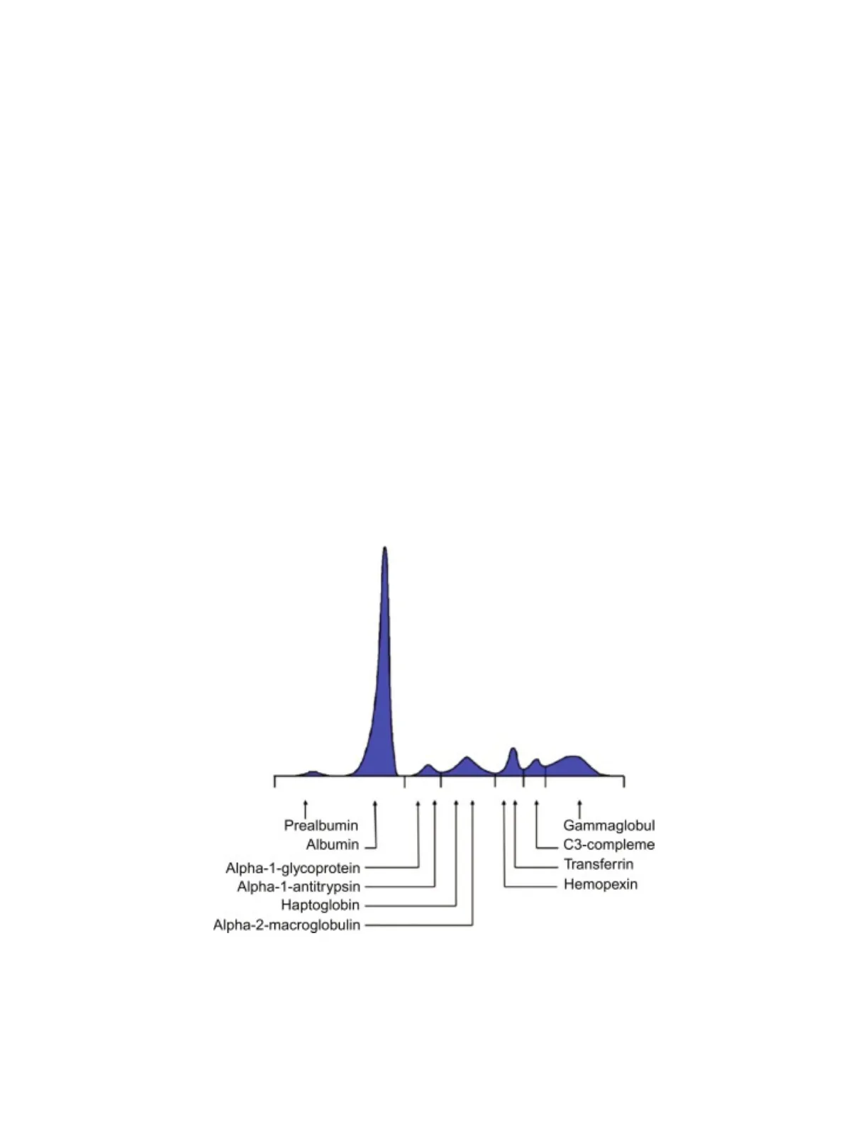
Prealbumin Region
Prealbumin (Transthyretin) Characteristics
- It is called prealbumin because it migrates faster than albumin (ALB) on protein electrophoresis.
- Serves as a transport protein for thyroid hormone thyroxine (T4) and retinol binding protein 4 (RBP4) in serum and CSF.
- Synthesized by both the liver and choroid plexus
- Negative APR protein (decreases during the acute phase)
- Protein is rapidly degraded during nutritional deficit and serves as a reservoir for these amino acids
- Used clinically as an indicator of nutritional status due to its rapid turnover and sensitivity to changes in protein synthesis and degradation.
İ Prealbumin Albumin Gammaglobul C3-compleme Transferrin Alpha-1-glycoprotein Alpha-1-antitrypsin Haptoglobin Hemopexin Alpha-2-macroglobulin Page 2
Albumin Region
Albumin (ALB) Properties
- (50 kDa) ALB has a strong negative charge at physiologic pH of 7.4
- The most abundant protein in plasma, (60%-65% of total protein)
- ALB functions as a carrier protein for various substances, including divalent cations, bilirubin, free fatty acids, and hormones such as thyroxine and estrogen, along with drugs such as penicillin, phenytoin, and warfarin
- ALB also functions as an amino acid reservoir
- The liver synthesizes ALB, and reduction of over 90% of the liver's functionality is necessary to decrease substantially the serum ALB concentrations
- In inflammatory disease, serum ALB decreases, because synthesis is inhibited by the cytokines interleukin-1 (IL-1) and interleukin-6 (IL-6), and/or tumor necrosis factor-a (TNF-a). (ALB is often referred to as a negative APR)
- Hyperalbuminemia= dehydration
- ALB normally is the most predominant protein excreted in urine, because it is the most abundant plasma protein and also because it is not entirely reabsorbed by the renal tubules.
Alpha-1 Region
Alpha-1 Antitrypsin (A1AT)
- al-Antitrypsin (A1AT): the main one in the al region.
- Inhibitor for trypsin, elastase, and several other proteases released by neutrophils
- Synthesized in the liver
- Robust positive APR protein that may function to attenuate proteases and other substances that are released during inflammation
Alpha-1 Acid Glycoprotein (AGP)
- al-Acid glycoprotein (AGP)
- Synthesized in the liver
- Is known to bind progesterone and other hormones, as well as drugs, such as lidocaine, propranolol, and phenobarbital Page 3
- Positive APR protein
Alpha-2 Region
Haptoglobin (Hp)
- Haptoglobin (Hp)
- Irreversibly binds free hemoglobin (Hb) in the blood (normally saturated). The resulting Hb-Hp complex is transported to the liver, where hemoglobin is broken down and its components (e.g., iron, globin) are recycled.
- Synthesized by hepatic parenchymal cells.
- Haptoglobin levels and hemolysis:
- Hp levels are normally high (hemoglobin levels are normally low).
- Decreased Hp levels indicate hemolysis because free hemoglobin levels increase and this saturates and depletes Hp quickly.
- Only a small amount of in vivo hemolysis is necessary to deplete Hp; for this reason, Hp measurements are clinically useful as a sensitive biomarker of intravascular hemolysis
Alpha-2 Macroglobulin (A2M)
- a2-Macroglobulin (A2M)
- Synthesized in the liver.
- One of the largest plasma proteins.
- Functions as an inhibitor and complexing agent for a broad spectrum of proteases (e.g., trypsin, thrombin, and plasmin).
- A2M concentrations are increased in the nephrotic syndrome because of induction of this protein's synthesis by the liver, presumably as a compensatory response to maintain oncotic pressure when ALB and other proteins are lost by urinary excretion.
- A2M protein is a negative APR protein (Note: this is what is written on the slides, but Alpha 2-macroglobulin is a positive APR according to google)
Ceruloplasmin (Cp)
- Ceruloplasmin (Cp)
Page 4
- Ferroxidase enzyme synthesized and catabolized by hepatic parenchymal cells
- Facilitate oxidation-reduction reactions in plasma such as the ferric (Fe III) to ferrous (Fe II) conversion of Fe-an essential part of Fe metabolism. It loads the iron in the ferritin.
- Stores and transports copper to the parts of the body that needs it. The transport is through the blood.
- Cp is a positive APR protein
- Cp can be used in screening for Wilson's disease
Wilson's Disease
A O Wilson's disease is 1 case per 30,000 live births in most populations (very rare). In the absence of this enzyme, copper is transported to the liver, but the metal is not subsequently bound to Cp. The result is the deposition of copper in hepatic parenchymal cells, in the brain, and in the periphery of the iris also known as "KayserFleischer" rings. KayserFleischer rings appear in approximately 95% of patients with this disorder.
Beta-1 Region
Transferrin (Tf)
- B1 Region: Transferrin (Tf)
- Binds and transports Fe in circulation
- Synthesized in the liver and reticuloendothelial system negative APR protein that is also decreased in chronic liver disease, malnutrition, renal insufficiency
- Transferrin is a negative acute-phase reactant (APR)
- Tf can be measured by immunoassay or estimated using its predictable relationship with total iron-binding capacity (TIBC) [TIBC= 1,43 x Tf (mg/dL). It is important to calculate the ability to transport and store iron. Page 5
- Approximately 70% of Tf binding sites are saturated with Fe, leaving excess capacity. In Fe deficiency anemia, the Fe concentration decreases, while the Tf protein and TIBC increase
Beta-2 Region
C3 Complement Protein
- ₿2 Region: C3
- C3 has the highest concentration of all complement proteins
- C3 synthesis occurs at several locations, including hepatic parenchymal cells (induced by cytokines and TNF-a), and by fibroblasts and monocytes (induced by endotoxin).
Gamma Region
Immunoglobulins in Gamma Electrophoretic Region
- y Region (most important): Immunoglobulin G (IgG), Immunoglobulin A (IgA),
Immunoglobulin M (IgM), Immunoglobulin D (IgD), and Immunoglobulin E (IgE)
represent the preponderance of protein in the y electrophoretic region
- IgG migrates to the gamma region (largest portion of immunoglobulins in circulation)
- IgA migrates to the beta region and the more anodal (early) gamma region
- IgM also tends to migrate toward the anodal portion of the gamma region
- Also, IgD and IgE but these are minority compared to the other three Igs.
- Immunoglobulins are synthesized mainly in lymphnodes and in blood by plasma cells
- In certain conditions (e.g., monoclonal gammopathies, such as multiple myeloma), a
paraprotein (or monoclonal antibody) may appear in this region.
- Paraproteins are antibodies that are produced abnormally, often by a single clone of plasma cells. These proteins can migrate in the gamma region, leading to abnormal peaks on electrophoresis.
Antigen binding site Light chain Fab fragment Disulfide bonds Heavy chain Fc fragment Page 6
Serum Protein Electrophoresis (SPEP)
Serum protein electrophoresis Anode Alb α. Output Scan Y Cathode
ALB and globulin proteins are two major protein groups in serum. The SPEP test separates these two protein groups on the basis of size and charge properties into the six major regions: ALB, a1, a2, B1, B2, and y.
After electrophoresis, the proteins are fixed in the gel Densitometry is performed with instrumentation that scans the SPEP Pattern. Software in the densitometer quantifies the proportion of total protein staining represented by each SPEP region.
UPEP (urine) is based on the same separation principles as SPEP. However, the concentration of protein in urine specimens is normally far lower than in serum; therefore preconcentration of samples is required before UPEP. Random or 24-hour urine specimens may be used in the analysis. Densitometric UPEP patterns are sometimes helpful for quantifying protein excretion and for monitoring the presence of monoclonal banding, particularly the excretion of k or 2 light chains in urine.
To understand if the liver and kidney are working compare serum and urine electrophoresis.
NORMAL PATTERN: Monoclonal protein not detected
Gel electrophoresis Albumin a1 a2 61 62 Y ! Albumin a-1 a-2 B-1 B-2 Y Low- resolution Gel Page 7
Can’t find what you’re looking for?
Explore more topics in the Algor library or create your own materials with AI.
