Tumori Odontogeni: Definizione, Classificazione Clinica e Ameloblastoma
Slide da Università sui Tumori Odontogeni. Il Pdf presenta una panoramica dettagliata sui tumori odontogeni, con particolare attenzione all'ameloblastoma, illustrando definizioni e classificazioni cliniche. Questo materiale didattico è utile per lo studio universitario della patologia orale.
Mostra di più54 pagine


Visualizza gratis il Pdf completo
Registrati per accedere all’intero documento e trasformarlo con l’AI.
Anteprima
Tumori Odontogeni: Definizione e Classificazione
Tumori Odontogeni Prof. Massimo PetruzziDEFINIZIONE Lesioni che derivano da elementi epiteliali (ectoderma) e/o mesenchimali (ectomesenchima) che partecipano alla formazione dell' elemento dentario.
Con il termine tumori odontogeni si fa quindi riferimento ad un gruppo di lesioni molto eterogeneo che ricomprende:
- Amartomi
- Tumori benigni
- Tumori maligni
Difficoltà Classificative dei Tumori Odontogeni
- Complessità dei tessuti coinvolti
- Estrema rarità di alcune lesioni, soprattutto quelle maligne
OM RDL V SR OEE IEE EM
Classificazione WHO-2022 dei Tumori Odontogeni
Tumori Odontogeni Benigni
Epitelio Odontogeno
- AMELOBLASTOMA
- Convenzionale
- Unicistico
- Extraosseo/periferico
- Adenoideo
- Metastatizzante
- TUMORE ODONTOGENO SQUAMOSO
- CARCINOMA ODONTOGENO A CELLULE CHIARE
- TUMORE ODONTOGENO EPITELIALE CALCIFICANTE (CEOT)
- CARCINOMA ODONTOGENO A CELLULE FANTASMA
- TUMORE ODONTOGENO ADENOMATOIDE
Epitelio + Ectomesenchima
- CARCINOSARCOMA ODONTOGENO
- FIBROMA AMELOBLASTICO
- TUMORE ODONTOGENO PRIMITIVO
- ODONTOMA
- -composto
- -complesso
- TUMORE DENTINOGENICO A CELLULE FANTASMA
Ectomesenchima
- SARCOMI ODONTOGENI
- FIBROMA ODONTOGENO
- MIXOMA ODONTOGENO
- CEMENTOBLASTOMA
- FIBROMA CEMENTO-OSSIFICANTE
Tumori Odontogeni Maligni
Carcinomi
- CARCINOMA AMELOBLASTICO
- CARCINOMA INRAOSSEO PRIMITIVO
- CARCINOMA ODONTOGENO SCLEROSANTE
Epidemiologia dei Tumori Odontogeni
COSTITUISCONO MENO DELL'1% DEI TUMORI CHE INTERESSANO IL CAVO ORALE
- < 2-3% di tutti i campioni inviati all'anatomo-patologo da chirurghi orali e maxillo-faciali
- 0.002-0.003% di tutti i tumori
- Si stima che i tumori benigni e gli amartomi abbiano un'incidenza di circa 100 volte maggiore rispetto ai maligni
- la maggior parte dei tumori odontogeni sono benigni ma alcuni di essi mostrano un'elevata aggressività locale e tendenza alla recidiva.
Eziologia: sconosciuta
Aspetti Clinici e Diagnostici dei Tumori Odontogeni
- Aspetto clinico: i benigni hanno crescita lenta ed espansiva con sintomatologia algica generalmente assente o molto lieve. Per contro il dolore e la rapida insorgenza di una tumefazione sono caratteristiche comuni a quasi tutti i maligni
- Imaging: data la compresenza di tessuti molli e duri l'aspetto può variare dalla radiotrasparenza alla radiopacità.
- Precursori: le cisti odontogene da sviluppo potrebbero essere coinvolte nel meccanismo di insorgenza di alcuni tumori odontogeni
Classificazione dei Tumori Odontogeni Benigni
- TUMORI BENIGNI CON SOLO EPITELIO ODONTOGENO
- TUMORI BENIGNI MISTI CON EPITELIO ED ECTOMESENCHIMA ODONTOGENO
- TUMORI BENIGNI CON SOLO ECTOMESENCHIMA ODONTOGENO
Ameloblastoma: Tumore Odontogeno Benigno Epiteliale
Definizione e Dati Epidemiologici dell'Ameloblastoma
AMELOBLASTOMA
- Definizione: Neoplasia odontogena benigna a crescita lenta, espansiva progressiva con tendenza ALLA RECIDIVA SE NON ADEGUATAMENTE RIMOSSA. Un tempo denominato ADAMATINOMA
- Dati epidemiologici
- 2º tumore odontogeno più comune (dopo l'odontoma)
- 0.5 casi anni su milione di abitanti.
- M=F
- Maggior incidenza: IV-V decade
- 80% mandibola (ANGOLO).
Istogenesi dell'Ameloblastoma
AMELOBLASTOMA
- Istogenesi: Residui della lamina dentaria(?) Residui del Malassez (?) Resti del Serres (?) Epitelio dell'organo dello smalto
- 90% dei casi mostra mutazioni dei geni della cascata MAPK
- Mutazioni nel Hedgehog (SHH) signaling pathways
Sandra F, Harada H, Nakamura N, Ohishi M. Midkine induced growth of ameloblastoma through MAPK and Akt pathways. Oral Oncol. 2004 Mar; 40(3):274-80. Wright JM, Vered M (2017) Update from the 4th edition of the World Health Organization classification of head and neck Tumours: odontogenic and maxillofacial bone tumors. Head Neck Pathol 11:68-77
Classificazione Clinica dell'Ameloblastoma
AMELOBLASTOMA
CLASSIFICAZIONE CLINICA
- Convenzionale (Multicistico) 85%
- Unicistico 14%
- Periferico 1%
- Metastatico
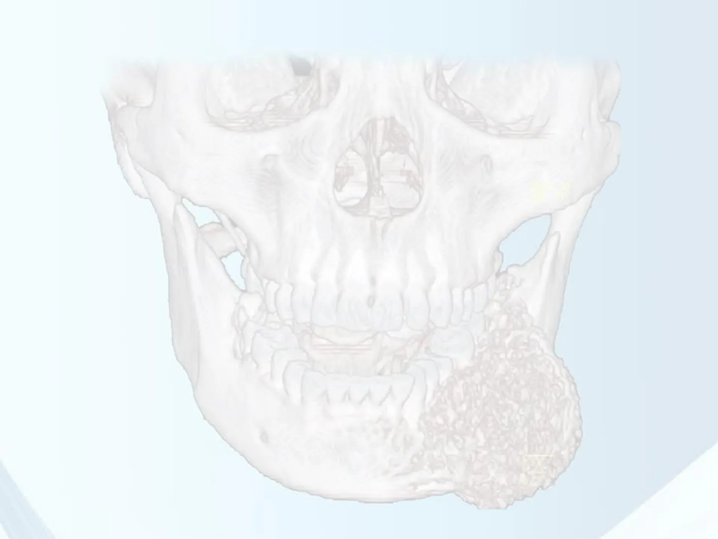
Ameloblastoma Convenzionale (Multicistico)
AMELOBLASTOMA
CLASSIFICAZIONE CLINICA
Convenzionale Noto anche come multicistico, per via dell'aspetto a «bolle di sapone» che radiologicamente mostra
- frequente Follicolare (Origina dall'epitelio dell'organo dello smalto) Plessiforme Acantomatoso A cellule granulose
CLASSIFICAZIONE istopatologica Nessuna implicazione prognostica
- Basaloide Desmoplastico Adenoideo
- frequente
Ameloblastoma Unicistico
AMELOBLASTOMA
CLASSIFICAZIONE CLINICA
Unicistico
- Area singola radiotrasparente, simile ad una cavità cistica.
- Diagnosticato nel 50% dei casi nella II decade di vita. Nell'80% dei casi si associa ad inclusione del III molare.
- Sede di insorgenza: più frequente alla mandibola in sede posteriore
- Può causare riassorbimento radicolare, corticale (1/3 dei casi),e non infiltra l'osso circostante
- Terapia: generalmente escisso con il sospetto di cisti follicolare; è bene considerare la possibilità di un ulteriore intervento in base alla profondità dell'invasione nella compagine della parete cistica. Possibili recidive anche dopo 10 anni
Crescita Componente Epiteliale nell'Ameloblastoma Unicistico
AMELOBLASTOMA
CLASSIFICAZIONE CLINICA
Unicistico Presenta un'unica area radiotrasparente (similcistica).
1 3 3 .2 In base alla crescita della componente epiteliale, l'ameloblastoma unicistico può essere suddiviso in:
- Luminale
- Intraluminale
- Intramurale
Maggior rischio di recidiva
Ameloblastoma Periferico (Extraosseo)
AMELOBLASTOMA
CLASSIFICAZIONE CLINICA
Periferico Detto anche extraosseo. Colpisce principalmente pazienti con un'età media di 52 anni e si verifica più frequentemente nella gengiva della mandibola. Raramente presenta recidive, anche se trattate in modo conservativo. L'aspetto istopatologico è rappresentato da isole di epitelio ameloblastico con un pattern simile al tipo convenzionale
RVG 6100
Ameloblastoma Metastatizzante
AMELOBLASTOMA
CLASSIFICAZIONE CLINICA
Metastatizzante
- Sebbene le metastasi non siano una caratteristica usuale della benignità, è stato segnalato che vari tumori benigni come l'adenoma pleomorfo, la sclerosi tuberosa e il tumore a cellule giganti dell'osso metastatizzano in sedi distanti. L'ameloblastoma può dare metastasi nonostante le caratteristiche istologiche benigne e questa variante è definita ameloblastoma metastatizzante.
Caratteristiche dell'Ameloblastoma Metastatizzante
AMELOBLASTOMA
CLASSIFICAZIONE CLINICA
Metastatizzante È stato classificato nella categoria dei tumori odontogeni maligni dall'OMS nel 2005, ma è stato riclassificato come tumore odontogeno epiteliali benigni nell'ultima classificazione dell'OMS del 2017. La ragione principale alla base del cambiamento è attribuita al fatto che gli ameloblastomi primari e metastatici sono istopatologicamente identici all'ameloblastoma « benigno» Sono stati descritti meno di 50 casi. Età media di insorgenza: 42 anni. È stata notata una leggera predilezione maschile. I casi mandibolari hanno mostrato più tendenza alla metastasi rispetto ai casi mascellari. L'ameloblastoma follicolare è stato riscontrato più frequentemente nel sito secondario seguito dal tipo plessiforme.
Pandiar D, Anand R, Kamboj M, Narwal A, Shameena PM, Devi A. Metastasizing Ameloblastoma: A 10 Year Clinicopathological Review with an Insight Into Pathogenesis. Head Neck Pathol. 2021 Jan 4.
Siti di Metastasi dell'Ameloblastoma
AMELOBLASTOMA
CLASSIFICAZIONE CLINICA
[Downloaded f Cas POLMONI (70%) Cavitating lung metastasis secondary to ameloblastoma Sir, Ameloblastoma are benign locally aggressive tumors accounting for about 1% of all oral tumors. Metastasis occurs in 2-5% of patients and lung is approximately involved in 80% of cases. Solitary or multiple lung nodules are the common manifestations, cavitating nodules have never been reported. Here we report probably the first case in literature about multiple cavitating lung secondaries in a 38-year-old female, who was previously diagnosed 5 years back to have ameloblastoma of right mandible. A 38-year-old female presented to pulmonary medicine outpatient department (CMC Vellore, Tamilnadu, India) with complaints of dry cough and exertional breathlessness of 3 months duration. There was no history of fever, loss of appetite, weight loss, hemoptysis, chest pain or symptoms suggestive of connective tissue disease (CTD) or vasculitis. Our patient had significant past history and 5 years back she was diagnosed to have benign ameloblastoma of right mandible [Figure 1a] and underwent total excision and repair with fibular graft [Figure 2a and b]. She was in regular follow-up and there was no signs of recurrence. Her general examination, systemic examination and laboratory investigations were within normal limits. Chest X-ray revealed [Figure 3a] multiple bilateral lung nodules with cavitation, and the same was confirmed by computed tomography [Figure 3b]. Her old chest X-ray taken 5 year back [Figure 3c] was normal. Her routine biochemistry and serology for CTD and vasculitis were negative. Bronchoscopy was done and the cultures were negative for mycobacteria and malignant cells were negative in cytology. Computed tomography (CT) guided transthoracic needle biopsy was done from one of the nodules and the histo Towed features oma [Figure 1b] suggestive of metastasizing amelo Local recurrence was ruled out with Mandibula radiographs. Metastasis to other with appropriate investiga ns. oncology department for fu her advised palliative chemotherapy, by her and was lost to follow-up. a b Figure 1: Mandibular surgical biopsy specimen (a) Showing characteristic features of benign ameloblastoma (hematoxylin-eosin; orginal magnification ×200) and transthoracic needle biopsy specimen (b) Showing features of metastasizing ameloblastoma (hematoxylin-eosin; original magnification x100) a b Figure 2: 3D volume reconstructed CT (a) Showing 4 x 3 cm expansile lytic lesion in the body of right mandible (black arrow) and post-reconstructed CT (b) Showing mandible defect reconstructed with fibular graft (white arrow) linfonodi loco regionali (28%) FEGATO Jo juinof sport Journal of Clinical Case Reports Case Report Open Access Metastatic Ameloblastoma to the Liver: Rare Presentation of a Rare Disease Lacin S1*, Dogrul A2, Dikmen E3, Kertmen N4, Turker A4 and Kars A4 'Department of Medical Oncology, Hacettepe Cancer Institute, Hacettepe University Sihhiye Campus, Oncology Hospital, Altındag, Turkey "Faculty of Medicine, Department of General Surgery, Hacettepe University, Turkey "Faculty of Medicine, Department of Thoracic Surgery, Hacettepe University, Turkey "Faculty of Medicine, Department of Medical Oncology, Hacettepe University, Turkey Abstract Ameloblastoma is a slow growing odontogenic epithelial neoplasm which originates from remnants of the dental lamina with a high recurrence rate, but a low tendency to metastasize. Locally invasive ameloblastoma is often aggressive and destructive, which erodes bone and invades adjacent structures. Despite a benign histology metastatic disease may occur and samples taken from metastatic tumor usually maintains the features of the original tumor. Ameloblastic carcinoma differs from ameloblastoma with malignant cytological features. Here we report an unusual case of ameloblastoma metastatic to lung and liver, unresponsive to systemic treatment with cisplatin and adriamycin, and well controlled with local surgical treatment. Keywords: Ameloblastoma; Metastasis; Liver; Resection Introduction Ameloblastoma is a rare, benign or cancerous tumor of odontogenic epithelium. Locally invasive ameloblastoma is often aggressive and destructive, which erodes bone and invades adjacent structures. While these tumors are rarely malignant and progress slowly. A 23-year-old man is presented in this case report. Case Report A 23-year-old man presented with three months history of increasing lower abdominal discomfort and a change in bowel habits. According to the hospital records, he was a nonsmoker and denied alcohol consumption. He had had dental problems in 2004, underwent dental extraction and operations then. Following these procedures, he had noticed a painless swelling in his right mandible. The lesion was resected, and histopathology was reported ameloblastomatous. Between 2004 and 2015, the patient suffered 7 local relapses which were treated surgically. In 2015the patient started to have the above mentioned complaints. CT scans of the chest and abdomen revealed two metastatic lesions in the rightlungand ahuge liver masslocated in segments 2 and 3 (Figures 1 and 2). A core biopsy taken from this lesion was reported as metastatic ameloblastoma. Immunohistochemistry revealed diffuse positive staining with cytokeratin (CK) 14, CK 19, CK 5, beta-catenin, focal staining with CK 18 and negative staining with calretinin. The patient received six cycles of cisplatin and adriamycin doublet. CT scans taken after the 3rd and 6th cycles showed stable disease. Despite these three additional cycles of this combination was given, without achieving an objective response. The patient was discussed in our multidisciplinary KARŞILAŞTIRMA TT Figure 1: Preoperative and after resection of the liver lesion abdominal CT scans view of the metastatic lesion in the liver. Medical Oncology, Hacettepe mpus, Oncology Hospital 2" Figure 3: (a) Chest X-ray PA view showing lung nodules with and without cavitations (white arrows), (b) same was seen on CT Thorax. (c) her old X ray taken 5 years back when she right mandibular ameloblastoma Metastasizing ameloblastoma ameloblastic carcinoma under the general grou Sopravvivenza a 5 anni 70% Figure 2: The patient thorax CT after resection of the pulmonary lesions. Received January 20, 2019; Accepted January 29, 2019; Published January 31, 2019 Citation: Lacin S, Dogrul A, Dikmen E, Kertmen N, Turker A, et al. (2019) Metastatic Ameloblastoma to the Liver: Rare Presentation of a Rare Disease. J Clin Case Rep 9: 1207. doi: 10.4172/2165-7920.10001207 Copyright: @ 2019 Lacin S, et al. This is an open-access article distributed under the terms of the Creative Commons Attribution License, which permits unrestricted use, distribution, and reproduction in any medium, provided the original author and source are credited. Lung India . Vol 32 . Issue 5 . Sep - Oct 2015 527 J Clin Case Rep, an open access journal ISSN: 2165-7920 Volume 9 . Issue 1 + 10001207 La diagnosi viene solitamente fatta in retrospettiva Ameloblastoma are uncommon slow-growing benign tumor arising from odontogenic epithelium of jaw. Usually occurs in middle-age group (20-40) and the most common site is mandible (80%). These tumors are locally aggressive and rarely metastasize. a C Metastatizzante Clinical Cas Lacin et al., J Cin Case Rep 2019, 9:1 DOI: 10.4172/2165-7920.10001207 ISSN: 2165-7101
Non hai trovato quello che cercavi?
Esplora altri argomenti nella Algor library o crea direttamente i tuoi materiali con l’AI.
