Special Senses: Visual System, Anatomy and Physiology of the Eye
Slides about Special Senses – Visual System. The Pdf explores the visual system, focusing on eye anatomy, vision physiology, and phototransduction. This university-level Biology presentation, produced in 2023, details structures like the retina, iris, and lens, along with the visual pathway, using diagrams and explanations for clarity.
See more30 Pages

Unlock the full PDF for free
Sign up to get full access to the document and start transforming it with AI.
Preview
Visual System Overview
Special Senses - Visual System In this lecture we will be covering ........
GERARD J. TORTORA BRYAN DERRICKSON PRINCIPLES OF ANATOMY & PHYSIOLOGY Organization, Support and Movement, and Control Systems of the Human Body 18TH PRITION VOLUM INTERNATIONAL STUDENT VERSION Chapter 17 Structure of the eye Physiology of vision Phototransduction The visual pathwayElectromagnetic spectrum
Electromagnetic Spectrum and Visible Light
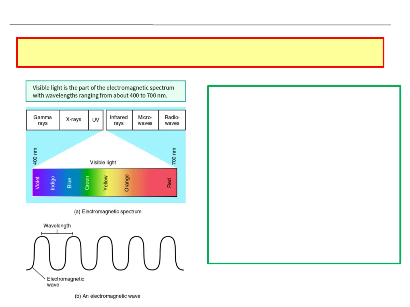
More than half the sensory receptors in the human body are located in the eyes, and a large part of the cerebral cortex is devoted to processing visual information Visible light is the part of the electromagnetic spectrum with wavelengths ranging from about 400 to 700 nm.
Gamma rays X-rays UV Infrared rays Micro- waves Radio- waves 400 nm 700 nm Visible light Violet Indigo Blue Green Yellow Orange Red (a) Electromagnetic spectrum Wavelength Electromagnetic wave
- An object can absorb certain wavelengths of visible light and reflect others; the object will appear the colour of the wavelength that is reflected
- A green apple appears green because it reflects mostly green light and absorbs most other wavelengths of visible light
- An object appears white because it reflects all wavelengths of visible light
- An object appears black because it absorbs all wavelengths of visible light (b) An electromagnetic wave
Accessory Structures of the Eye
The accessory structures of the eye include the eyelids, eyelashes, eyebrows, the lacrimal (tear-producing) apparatus, and extraocular muscles
The palpebral fissure is the space between the upper and lower eyelids that exposes the eyeball.
Eyebrow Pupil (under cornea) Iris Upper eyelid Lacrimal caruncle Eyelash George Diebold/Getty Images Lower eyelid Medial commissure Lateral commissure Conjunctiva (over sclera) Palpebral fissure
Eyelids and Protection
Eyelids (palpebra) shade the eyes during sleep, protect the eyes from excessive light and foreign objects, and spread lubricating secretions over the eyeballs
In the medial commissure is a small, reddish elevation, the lacrimal caruncle which contains sebaceous glands and sudoriferous glands
Sclera - "white" of the eye Cornea - transparent region that forms the outer anterior surface of the eyeball
Eyelashes/eyebrows - help protect the eyeballs from foreign objects, perspiration, and the sun
Infection in sebaceous glands at the base of the eyelash hair follicles - a sty.
Accessory Structures of the Eye Muscles
The accessory structures of the eye include the eyelids, eyelashes, eyebrows, the lacrimal (tear-producing) apparatus, and extraocular muscles
Levator palpebrae superioris muscle Superior rectus muscle Orbicularis oculi muscle Eyebrow Bulbar conjunctiva Palpebral conjunctiva Scleral venous sinus Cornea Optic nerve Pupil Lens Iris Upper eyelid Eyelashes Lower eyelid Tarsal glands Inferior oblique muscle Orbicularis oculi muscle Inferior rectus muscle
- Eyes sit in the bony depressions of the skull called the orbits
- Six extraocular muscles extend from the walls of the bony orbit to the sclera
- Surrounded by periorbital fat
- Muscles are capable of moving the eye in almost any direction
- Motor units in these muscles are small - two or three muscle fibres per neuron
Lacrimal Apparatus of the Eye
Group of structures that produces and drains lacrimal fluid or tears in a process called lacrimation
Flow of Tears
FLOW OF TEARS Upper eyelid Lacrimal gland secretes tears into Superior lacrimal canaliculus Lacrimal gland Lacrimal puncta Excretory lacrimal duct Excretory lacrimal sac Inferior lacrimal canaliculus Superior or inferior lacrimal canaliculi drain tears into Lower eyelid Excretory nasolacrimal duct Lacrimal sac, which drains tears into Inferior nasal concha Nasal cavity Nasolacrimal duct, which drains tears into Nasal cavity (b) Anterior view of the lacrimal apparatus
- Lacrimal fluid is a watery solution containing salts, some mucus, and lysozyme, a protective bactericidal enzyme - protects, cleans, lubricates, and moistens the eyeball
- Lacrimation is a protective mechanism, as the tears dilute and wash away the irritating substance Excretory lacrimal ducts, which distribute tears over surface of eyeball
Anatomy of the Eyeball - Fibrous Tunic
The wall of the eyeball consists of three layers: the fibrous layer, the vascular layer, and the inner layer (retina).
Composed of two parts: 1) Anterior cornea 2) Posterior sclera
Transverse plane Anterior segment of eyeball (contains aqueous humor): Light Anterior chamber Visual axis Cornea Posterior chamber Pupil Iris Lens Zonular fibers Lacrimal sac Bulbar conjunctiva Ciliary body: Ciliary muscle Ciliary process Ora serrata Retina Choroid Hyaloid canal Sclera Medial rectus muscle Lateral rectus muscle Posterior segment (contains vitreous humor) Macula MEDIAL LATERAL Central retinal artery and vein Optic (II) nerve Optic disc Fovea centralis Scleral venous sinus
Cornea and Sclera
O Cornea is a transparent coat that covers the coloured iris As it is curved, the cornea helps focus light onto the retina Sclera = the "white" of the eye, is a layer of dense connective tissue made up mostly of collagen fibres and fibroblasts Covers the entire eyeball except the cornea; it gives shape to the eyeball, and serves as a site of attachment for the extraocular muscles
Anatomy of the Eyeball - Vascular Tunic
The wall of the eyeball consists of three layers: the fibrous layer, the vascular layer, and the inner layer (retina).
Composed of three parts:
Transverse plane Anterior segment of eyeball (contains aqueous humor): Light Anterior chamber Visual axis Cornea Posterior chamber Iris Lens Zonular fibers Lacrimal sac Bulbar conjunctiva Ciliary body: Ciliary muscle Ciliary process Ora serrata Retina Choroid Hyaloid canal Sclera Medial rectus muscle Lateral rectus muscle Posterior segment (contains vitreous humor) Macula MEDIAL LATERAL Central retinal artery and vein Optic (II) nerve Optic disc Fovea centralis 1) Choroid 2) Ciliary body 3) Iris Scleral venous sinus Pu
Choroid and Ciliary Body
- Choroid
- highly vascularized - provides nutrients to the posterior surface of the retina
- contains melanocytes - produce melanin - absorbs stray light rays, prevents reflection and scattering of light within the eyeball - images remain sharp and clear
- Ciliary Body
- Ciliary processes are protrusions or folds - zonular fibres or suspensory ligaments extend from the ciliary process and attach to the lens
- Ciliary muscle is a circular band of smooth muscle. Contraction or relaxation changes the tightness of the zonular fibres, which alters the shape of the lens, adapting it for near or far vision
Iris and Pupil Regulation
- Iris
- coloured part of eyeball suspended between the cornea and the lens
- amount of melanin in the iris determines the eye colour
- regulates the amount of light entering the eyeball through the pupil
- autonomic reflexes regulate pupil diameter in response to light levels Contraction of the sphincter pupillae muscles causes constriction of the pupil; contraction of the dilator pupillae causes dilation of the pupil.
Pupil constricts as sphincter pupillae muscles of iris contract (parasympathetic) Pupil Pupil dilates as dilator pupillae muscles of iris contract (sympathetic) Visuals Unlimited, Inc./Dr. Richard Kessel & Dr. Randy Kardon/Tissues & Organs/Getty Images Iris Pupil Ciliary process Bright light Normal light Dim light SEM 15x (b) Posterior surface of iris and ciliary process, lens removed Anterior views
Anatomy of the Eyeball - Retina
The wall of the eyeball consists of three layers: the fibrous layer, the vascular layer, and the inner layer (retina).
Transverse plane Anterior segment of eyeball (contains aqueous humor): Light Anterior chamber Visual axis Cornea Posterior chamber Iris Lens Zonular fibers Lacrimal sac Bulbar conjunctiva Ciliary body: Ciliary muscle Ciliary process Ora serrata Retina Choroid Hyaloid canal Sclera Medial rectus muscle Lateral rectus muscle Posterior segment (contains vitreous humor) Macula MEDIAL LATERAL Central retinal artery and vein Optic disc Fovea centralis Optic (II) nerve Pupil Scleral venous sinus
Retina Structure and Visual Acuity
Retina
- . lines the posterior three-quarters of the eyeball and is the beginning of the visual pathway The optic disc is the site where the optic nerve exits the eyeball. The fovea centralis is the area of highest visual acuity.
NASAL SIDE TEMPORAL SIDE Macula Optic disc Retinal blood vessels Fovea centralis Paul Parker/Science Source Left eye Examined for pathological changes, such as hypertension, diabetes mellitus, cataracts, and age-related macular disease Several landmarks are visible through an ophthalmoscope;
- Optic disc (blind spot)
- site where the optic nerve exits the eyeball Retinal blood vessels
- Macula
- exact centre of posterior retina
- Fovea centralis
- small depression in centre of macula contains only cones
- area of highest visual acuity/resolution (sharpness)
Microscopic Structure of the Retina
- Sclera Choroid Pigmented layer of the retina Photoreceptor cell layer (rods and cones) Outer synaptic layer Bipolar cell layer Neural layer of the retina Mark Nielsen Inner synaptic layer Ganglion cell layer Optic (II) nerve axons LM 280x Central retinal artery Central retinal vein Optic (II) nerve Sclera Optic disc (blind spot) 0 Choroid Pigmented layer of the retina Neural layer of the retina (b) Transverse section of posterior eyeball at optic disc
Consists of TWO layers 1) Pigmented layer (contains melanin) 2) Neural (sensory) layer - multi-layered outgrowth of brain - processes visual data before sending to optic nerve
Photoreceptors: Rods and Cones
Outer segments Pigmented layer of the retina Rod Inner segments Cone Photoreceptor cell layer - Outer synaptic layer Horizontal cell Bipolar cell Bipolar cell layer Neural layer of the retina Amacrine cell Inner synaptic layer Synaptic terminals Ganglion cell Ganglion cell layer Optic (II) nerve axons Retinal blood vessel Direction of nerve impulses through retina Direction of incoming light Nerve impulses propagate along optic nerve axons toward optic disc (a) Microscopic structure of the retina
- There are two types of photoreceptors: rods (~120 million) and cones (~6 million)
- Rods allow us to see in dim light - do not provide colour vision - so in dim light we can see only black, white, and all shades of grey
- Brighter lights stimulate cones, which produce colour vision; (1) blue cones, (2) green cones, and (3) red cones
- Colour vision results from the stimulation of various combinations of these cones
Can’t find what you’re looking for?
Explore more topics in the Algor library or create your own materials with AI.
