Unit 6 Lesson 2: Hypersensitivity in general pathology
Slides from Ucam about Unit 6 Lesson 2. Hypersensitivity. The Pdf, a presentation for University-level Biology students, details hypersensitivity, its classification, and mechanisms, with a focus on Type I, including mediators and cells involved, using diagrams and comparative tables.
See more31 Pages


Unlock the full PDF for free
Sign up to get full access to the document and start transforming it with AI.
Preview
UCAM Unit 6 Lesson 2: Hypersensitivity
UCAM Unit 6 Lesson 2. Hypersensitivity. General Pathology Begoña Alburquerque González, PhD. Grade in Veterinary Medicine.
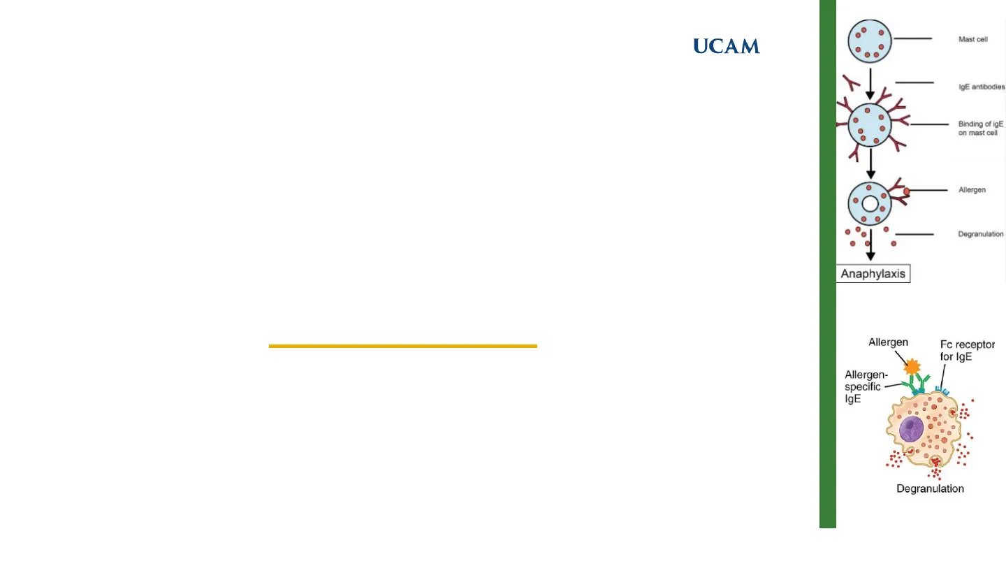
Mast cell IgE antibodies Binding of igE on mast cell Allergen Degranulation Anaphylaxis Allergen Fc receptor for IgE Allergen- specific IgE DegranulationUCAM
Contents Overview
CONTENTS ❑ Introduction - Hypersensitivity: ✓ Type I. ✓ Type II. ✓ Type III. ✓ Type IV. 2UCAM
Introduction to Hypersensitivity
Introduction Hypersensitivity V It refers to a situation of abnormal reactivity, in which the organism reacts with an exaggerated or inappropriate immune response to something that it perceives as a foreign substance. Disorders caused by immune responses are called Hypersensitivity Disorders.
ADCC Allergen Fc receptor for IgE Fc receptor Cytotoxic cell Allergen- specific IgE Surface Target antigen cell Complement activation Neutrophil Immune complex Degranulation Activated macrophage
Types of Hypersensitivity Reactions
Type Type II Type III Type IV IgE-Mediated Hypersensitivity IgG-Mediated Cytotoxic Hypersensitivity / 1 9M Immune Complex-Mediated Hypersensitivity Ag - Ab Cell-Mediated Hypersensitivity TCDS - TEDY
- Ag induces crosslinking of IgE bound to mast cells and basophils with release of vasoactive mediators
- Ab directed against cell surface antigens meditates cell destruction via complement activation or ADCC
- Ag-Ab complexes deposited in various tissues induce complement activation and an ensuing inflammatory response mediated by massive infiltration of neutrophils
- Sensitized Ty1 cells release cytokines that activate macrophages or Tc cells which mediate direct cellular damage
Manifestations of Hypersensitivity
- Typical manifestations include systemic anaphylaxis and localized anaphylaxis such as hay fever, asthma, hives, food allergies, and eczema
- Typical manifestations include blood transfusion reactions, erythroblastosis fetalis, and autoimmune hemolytic anemia
- Typical manifestations include localized Arthus reaction and generalized reactions such as serum sickness, necrotizing vasculitis, glomerulnephritis, rheumatoid arthritis, and systemic lupus erythematosus
·Antigen Immune complex C36) C3b Sensitized TDTH C3b Complement activation Cytokines 6003
- Typical manifestations include contact dermatitis, tubercular lesions and graft rejection 3Introduction
Hypersensitivity Characteristics
UCAM Hypersensitivity C Varies in severity Chronic, debilitating Any age, any gender 20-40 years, more women Affects 25% population 2 Affects 2-5% population O Allergic Diseases Autoimmune Diseases Comparing allergic and autoimmune diseases. 4UCAM
Hypersensitivity Type I: IgE-Mediated Reactions
Hypersensitivity Type I V They are mediated by (IgE) antibodies produced in response to allergens: "harmless" antigens. V The antigens or allergens capable of inducing this type of reaction are pollen proteins, dust mites, animal epithelia and chemicals such as penicillin,) The immediate name is due to the fact that it takes a few minutes for the symptoms to start once the reaction between the allergen and the IgE antibody occurs. There is a strong genetic predisposition for the development of allergic diseases. This predisposition is called atopy.
Allergen CD4 IL-4 B cell TH cell + Innate Allergen Small blood vessel Vasoactive Fc receptor for IgE amines ** Mucous gland Blood platelets + Allergen Memory cell Plasma cell Sensitized mast cell Degranulation Sensory-nerve endings Allergen- specific IgE receptors in ( the surface for 1g E Eosinophil binding directly to most well ( not to Beel !) Type I Hypersensitive Reaction Sequence 1 Allergen Exposure IgE Production Second Allergen Exposure Mediator Release Increased Vascular Permeability O -O B Cell [Activation IgE Binding to Mast Cells IgE Crosslinking Smooth Muscle Contraction innate , adaptative ) ? "Smooth muscle cellUCAM
Hypersensitivity Type I: Cells Involved
Hypersensitivity Type I name of the Fc receptor - FcERI ( for Ig f )
Mast Cells, Basophils, and Eosinophils
MAST CELLS BASOPHILS EOSINOPHILS
| Characteristic | Mast Cells | Basophils | Eosinophils |
|---|---|---|---|
| Major site of maturation | Bone marrow precursors mature in connective tissue and mucosal tissues | Bone marrow | Bone marrow |
| Location of cells | Connective tissue and mucosal tissues | Blood (~0.5% of blood leukocytes); recruited into tissues | Blood (~2% of blood leukocytes); recruited into tissues |
| Life span | Weeks to months | Days | Days to weeks |
| Major growth and differentiation factor (cytokines) | Stem cell factor, IL-3 | IL-3 | IL-5 |
| Expression of FcERI | High | High | Low |
| Major granule contents | Histamine, heparin and/or chondroitin sulfate, proteases | Histamine, chondroitin sulfate, protease | Major basic protein, eosinophil cationic protein, peroxidases, hydrolases, lysophospholipase |
FcERI, Fce receptor type 1; IL, interleukin. 6UCAM
Hypersensitivity Type I: FcERI Receptor and Signaling
Hypersensitivity Type I . a chain , involved in the binding to the Fc portion of the heavy chain & of IgE . The @ chain and the y chains mediate signal transduction via ITAM (immunoreceptor tyrosine-based activation motif domains). Bound IgE antibody FLERA receptor Y Y NN B
Effects of IgE Binding on Cell Types
Cell Types 3G Effects of IgE Binding Mast Cells Basophils - Increased Receptor Expression Eosinophils Promotes Cell Survival Dendritic Cells
FcERI Receptor a Chain ß Chain Binding to Fc Portion Signal Transduction of the union between Igt and receptor Y Chains ITAM Domains granulation due to phosphorilation- cell are going to receive signal to granulation 3 # domains contains 3# domains Syk P P Lyn P P P N C C / C ITAMs + it activate 1 produce C 73 Ag = Allergen Bcell activating the releasing of granulocytes FLERI - Igf granules released by receptor of FLERI P ITAM domain _ 3 subunits 8 - @ MAST CELL a + (Bind to Antibody) (containing the receptenUCAM
Hypersensitivity Type I: Degranulation and Activation
Hypersensitivity Type I Antigen IgE IgE Fc receptor Signals for degranulation Signals for activation of phospholipase A2 · Histamine - Granule contents · Proteases Immediate response Vasodilation Vascular leakage Smooth muscle spasm · Chemotactic factors (ECF, NCF) Mast cell PAF Membrane phospholipids Prostaglandin D2 Nucleus Arachidonic acid Leukotrienes B4, C4, D4 Late-phase reaction Leukocyte infiltration Epithelial damage Bronchospasm Signals for cytokine gene activation Secreted cytokines Figure 5.18 Degranulation and Activation of Mast Cells. When an animal is sensitized by antigen in a type I hypersensitivity reaction, it has immunoglobu- lin E (IgE) molecules bound to receptors on the surface of mast cells. When exposed to antigen, the bound IgE on the mast cell surface is cross-linked, resulting in activation of the mast cell. This leads to degranulation of preformed mediators and the synthesis of mediators largely though activation of phospholipase A2, which lead to inflammatory responses. ECF, Eosinophil chemotactic factor; NCF, neutrophil chemotactic factor; PAF, platelet-activating factor. 8UCAM
Hypersensitivity Type I: Mediator Effects
Hypersensitivity Type I Induce Vascular Leakage Vasoactive Amines Bronchocon striction Vascular leak Vasoactive amines (e.g., histamine) Broncho- constriction Lipid mediators (e.g., PAF, PGD2, LTC4) Cytokines Mast Cells and Basophils > Contribute to Inflammation Lipid Mediators Intestinal hypermotility Activated mast cell (or basophil) Cytokines (e.g., TNF) Inflammation Lipid mediators (e.g., PAF, PGD2, LTC4) Enzymes (e.g., tryptase) Tissue damage 9 Eosinophils Toxic to Parasites Cationic granule proteins (e.g., major basic protein, eosinophil cationic protein) Killing of parasites and host cells Eosinophil Enzymes (e.g., eosinophil peroxidase) Tissue damage Enzymes Contribute to Tissue Damage Enzymes e Cationic Proteins Toxic to Host Cells 9 Vasodilation Intestinal HypermotilityUCAM
Hypersensitivity Type I: Reaction Phases
Hypersensitivity Type I Immediate Late phase reaction A B C Allergen exposure MAST CELLS Eosinophils Neutrophils Lymphocytes 2 Mast cell degranulation Clinical manifestations Edema Vascular congestion Eosinophils 0 1 4 8 12 16 20 Hours after allergen exposure Immediate Hypersensitivity Reaction. A, Early reaction (minutes) is characterized by mast cell degranulation and release of preformed vasoactive substances that cause vasodilation and increased vascular permeability, resulting in edema of interstitial tissue. B, As the lesion progresses to the late phase (hours), the inflammatory infiltrate is primarily composed of eosinophils and fewer lymphocytes and neutrophils. (A and B courtesy Dr. Daniel Friend, Department of Pathology, Brigham and Women's Hospital, Boston.) 10UCAM
Hypersensitivity Type I: Pathogenesis
Hypersensitivity Type I B lymphocyte Exposure to allergen TH2 cell Activation of TH2 cells and IgE class switching in B lymphocytes Allergen (e.g., pollen) Mucosal lining IgE-secreting B lymphocyte Production of IgE IgE FcERI Mast cell Binding of IgE to FcERI on mast cells 2) Repeat exposure to allergen Immediate hypersensitivity reaction (minutes after repeat exposure to allergen) Vasoactive - amines, lipid mediators Activation of mast cell: release of mediators Mediators Late-phase reaction (2-8 hours after repeat exposure to allergen) Cytokines Type I Hypersensitivity Reaction. The pathogenesis of a type I hypersensitivity reaction begins with exposure to antigen (allergen) that results in activation of T helper lymphocyte type 2 (TH2) lymphocytes and B lymphocytes, leading to the production of immunoglobulin E (IgE) and the sensitizing of mast cells. Continued or repeat exposure to antigen results in cross- linking of IgE bound to mast cells, causing activation and release of inflammatory mediators. 11UCAM
Hypersensitivity Type I: Disorders
Hypersensitivity Type I : Disorders How to address allergic reactions in pets?
- Canine atopic dermatitis Allergic skin reaction in dogs due to environmental allergens.
- Feline asthma Hypersensitivity reaction in cats causing airway inflammation and respiratory distress.
- Anaphylaxis in dogs and horses Severe allergic reaction to allergens like vaccines, insect stings, or certain foods. 12UCAM
Hypersensitivity Type I: Diagnosis
Hypersensitivity Type I : Treatment Clinical History and Physical Examination > Type of Test?
- Skin Tests
- Provocation Tests
- Quantification of Total IgE Levels
- Quantification of Specific IgE
- Basophil Activation Test
- Measurement of Inflammatory Cell Markers
- Patient Sensitization Profile
- Mastocytic Triptase
- Cationic Eosinophilic Protein 13UCAM
Hypersensitivity Type I: Treatment Strategies
Hypersensitivity Type I 0 Prevent contact with the allergen that triggers the disease. " The use of drugs that are directed to inhibit degranulation of the mast cell, to antagonize the effects of mediators of mast cells and to reduce inflammation. Immunotherapy (allergenic vaccines).
| Syndrome | Therapy | Mechanism of action |
|---|---|---|
| Anaphylaxis | Epinephrine | Causes vascular smooth muscle contraction; increases cardiac output (to counter shock); inhibits further mast cell degranulation |
| Bronchial asthma | Corticosteroids Leukotriene antagonists Phosphodiesterase inhibitors | Reduce inflammation Reduce inflammation Relax bronchial smooth muscles |
| Various allergic diseases | "Desensitization" (repeated administration of low doses of allergens) AntilgE antibody Antihistamines Cromolyn | Unknown; may inhibit IgE production and increase production of other lg isotypes; may induce T cell tolerance Neutralize and eliminate IgE Block actions of histamine on vessels and smooth muscles Inhibits mast cell degranulation |
Can’t find what you’re looking for?
Explore more topics in the Algor library or create your own materials with AI.
