Oral Mucosa and Buccopharyngeal Lymphatic Ring, UCAM Presentation
Slides from Ucam Universidad Católica De Murcia about Oral Mucosa and Buccopharyngeal Lymphatic Ring. The Pdf provides a detailed overview of the histology of the oral mucosa and the buccopharyngeal lymphatic ring, including the basement membrane and lamina propria, suitable for university Biology students.
See more39 Pages


Unlock the full PDF for free
Sign up to get full access to the document and start transforming it with AI.
Preview
Oral Mucosa and Buccopharyngeal Lymphatic Ring
Histology
Ana Isabel García Guillén, Ph.D. Bachelor in Dentistry
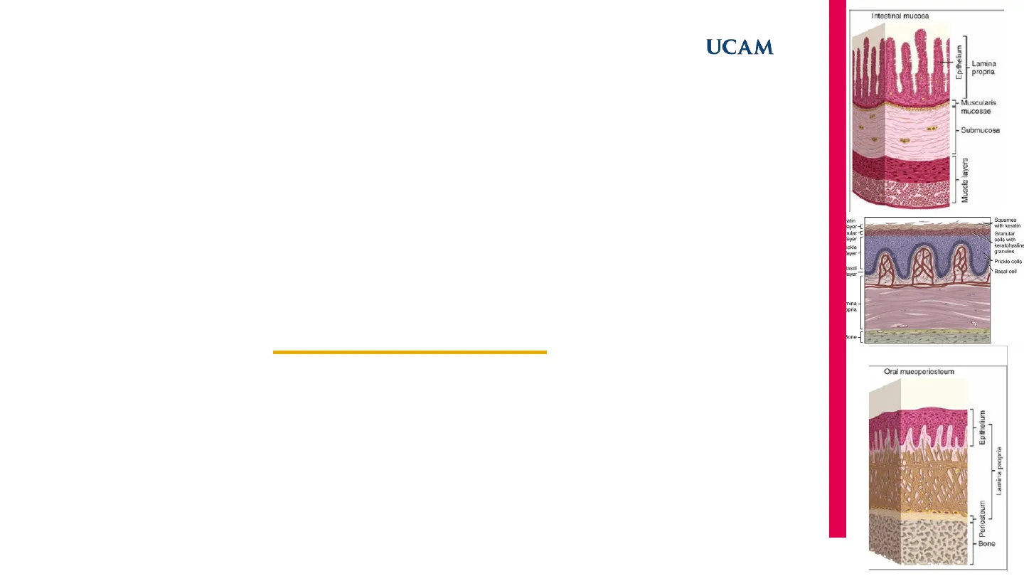
Intestinal Mucosa
Epithelium Lamina propria - Muscularis mucosae - Submucosa JL Muscle layers latin ayer -[ ular -C laye Ickle ayer- asal aye mina . bpria Bone
Oral Mucoperiosteum
Squames with keratin Granular cells with keratohyaline granules Prickle cells Basal cell Epithelium Lamina propria Periosteum - BoneUCAM
Oral Mucosa
UCAM Universidad Católica de Murcia C UCAMUCAM
Oral Cavity
Introduction to Oral Cavity Structures
- Mouth is an oval-shaped cavity inside the skull.
- Functions: eating and speaking.
Main Parts of the Oral Cavity
- Lips
- Cheecks
- Vestibule
- Gums
- Hard and soft palate
- Teeth
- Tongue
- Salivary glands.
- Tonsils For another unit 3UCAM
Oral Cavity Mucosa Types
Lining mucosa Gingival crevice Buccal mucosa Tooth Bacterial biofilm Vessel Masticatory mucosa Crevicular epithelium Junctional epithelium Tongue mucosa Keratin layer > porsum of tounge Trends in Immunology Oral Cavity
Oral Mucosa Epithelium
> Epithelium BCT · Surface of the oral cavity · It is a mucous membrane. Form by: . Stratified squamous epithelium (can be keratinized) Gingiva Hard palate 1 · Connective tissue termed lamina propria. . Continuous with the skin of the lip through the vermilion border. Ly search up . Posteriorly, with the mucosa of the pharynx. . Attached by a loose connective tissue termed submucosa to the underlying structure which may be bone or muscle > Not always present 4 Floor of mouth Keratin layer Bacteria Tooth -Junctional epitheliumUCAM
Oral Mucosa Structure and Classification
· Structure varies and adapts to function in different regions of the oral cavity. · Classification: Functional criteria Type of epithelium
Masticatory Mucosa
Masticatory mucosa (gingiva and hard palate) > Hard Structures that have mechanical Stress : Keratinized KERATINIZED All of these places don't have hydration Issures or a hard structure (2)
Lining or Reflecting Mucosa
Lining or reflecting mucosa (lip, cheek, vestibular fornix, alveolar mucosa, floor of mouth and soft palate) NON-KERATINIZED
Specialized Mucosa
Specialized mucosa (dorsum of the tongue and taste buds) KERATINIZED 5UCAM
Functions of the Oral Mucosa
· Defense or protection · Separates and protect deeper tissues from oral environment . Show adaptations of the epithelium to mechanical forces (compression, stretching, abrasions) · Barrier to microorganisms · Lubrication or secretion · Saliva (salivary glands) . Maintain the oral cavity moist and prevent drying. · Sensory · Temperature, touch and pain. . Taste is only in the tongue (taste buds) 6UCAM
Oral Mucosa Composition
The two main tissue components: • Oral epithelium stratified squamous epithelium · Lamina propria underlying connective tissue layer The interface between epithelium and connective tissue, connective tissue papillae · usually irregular · upward projections of connective tissue that interdigitates with epithelial ridges or pegs There is a basement membrane at the interface between epithelium and connective tissue - Epithelium - Lamina propria - Submucosa Periosteum Bone V 7UCAM
Oral Mucosa Composition Comparison
Intestinal mucosa Oral mucosa Oral mucoperiosteum Epithelium Lamina propria Epithelium Epithelium L J- Muscularis mucosae Lamina propria - Submucosa Lamina propria T Muscle layers Submucosa Periosteum - Bone Bone A B C Note: Mucoperiosteum: In regions such as the gingiva and parts of the hard palate, oral mucosa is attached directly to the periosteum of underlying bone, with no intervening submucosa. It is a firm, inelastic attachment. 8UCAM
Oral Epithelium Stratified Squamous Epithelium
· Keratinized · Orthokeratinized: gingiva and hard palate · Parakeratinized: some parts of hard palate and gingiva- surface layer has keratin but nuclei are retained in many of the squames (pycknotic nuclei) · Non-keratinized: cheek, sublingual tissues, soft palate, ... Pyknotic nuclei. Keratinized Keratinized layer Granular layer layer Granular layer Superficial layer Intermediate layer Prickle cell layer Prickle cell layer Prickle cell layer Basal cell Player Basal cell layer Basal cell layer 50 um A B C A, Orthokeratinization in human gingiva. Nuclei are lost in the keratinized surface layer. B, Parakeratinization in human gingiva. The keratin squames retain their pyknotic nuclei (low cellular metabolism) C, Non-keratinization in primate buccal epithelium. No clear division of strata exists, and nuclei are apparent in the surface layer. 9UCAM
Keratinized Epithelium Layers
(inflexible, tough, resistant to abrasion, and tightly bound to the lamina propria) 4 layers: • Basal layer (stratum basale) • Spinous (prickle) layer (stratum spinosum) • Granular layer (stratum granulosum) • Cornified layer (stratum corneum)
Basal Layer (Stratum Basale)
· Basal layer (stratum basale) . 1 layer of cuboidal or columnar cells adjacent to the basal lamina (basement membrane) · Undergo mitosis, thus providing new cells (Ki67+) · Specialized structures called hemidesmosomes, that connect with the basement membrane and lateral desmosomes (with numerous tonofilaments) Keratin filaments [tonofilaments) Keratin intermediate filaments (tonofilaments) Plate Plaque Plasma membrane Basal lamina Plectir BPAG1 (bullous pemphigoid antigen 1) 230 kd BPAG2 (bulous Integrin subunit pemphigoid B4 integrin antigen 2) 180 kd Anchoring filament (laminin 5) Plaque Plate Anchoring filaments Basal lamina Hemidesmosomes BPAG1 (a member of the plakin family) and BPAG2 (a transmembrane protein with an extracelular collagenous domain) connect the basal lamina to intermediate filaments. Plectin (a member of the plakin family) and integrin subunit ß4 (forming a complex with integrin subunit og) link the basal lamina to intermediate filaments. Kierszenbaum & Tres: Histology and Cell Biology: An Introduction to Pathology, 3e Copyright @ 2012 by Mosby, an imprint of Elsevier Inc. All rights reserved. Keratin layer -[ Granular -C layer Squames with keratin Prickle layer - Granular cells with keratohyaline granules Prickle cells Basal layer Basal cell Lamina propria Bone - Orthokeratinized Stratified Squamous Epithelium (and deeper tissues) 10UCAM
Spinous (Prickle) Layer
Keratinized epithelium (inflexible, tough, resistant to abrasion, and tightly bound to the lamina propria) 4 layers: · Spinous (prickle) layer (stratum spinosum) . Some layers of larger elliptical or spherical cells joining by desmosomes (On the basis of light microscope, appears that the cells present intercellular spaces and are joined by 'intercellular bridges'- gives the name) . Intermedial cells that present tonofilaments of keratin. Keratin Squames with keratin layer -[ Granular -C layer Granular cells with keratohyaline granules Prickle layer - Prickle cells Basal layer Basal cell Lamina - propria Bone Orthokeratinized Stratified Squamous Epithelium (and deeper tissues) Tonofilaments (keratin-containing intermediate filaments) Desmosomal plaque (desmoplakin) Cadherins Spinelike cytoplasmic processes Melanocyte Dermis Melanin granules transferred to keratinocytes Desmosomes link the cytoplasmic processes (or spines) of adjacent keratinocytes Keratinocytes in the stratum spinosum are seen in a tangential section of the epidermis. Note the spinelike cytoplasmic processes of the keratinocytes. These processes contain bundles of tonofilaments inserted in the plaques of desmosomes linking cell processos derived from adjacent keratinocytos. Kierszenbaum & Tres: Histology and Cell Biology: An Introduction to Pathology, 3e Copyright @ 2012 by Mosby, an imprint of Elsevier Inc. All rights reserved. 11Oral Mucosa
Granular Layer (Stratum Granulosum)
Keratinized epithelium 4 layers: • Granular layer (stratum granulosum) . Larger flattened cells containing small basophilic granules · Granules are called keratohyalin granules . Epidermal and oral keratinocytes express additional differentiation markers, including filaggrin and trichohyalin, that associate with the keratin cytoskeleton during terminal differentiation. . Protein synthesis rate diminishes as the cell approaches the stratum corneum UCAM Keratin Squames with keratin layer -[ Granular -C layer Granular cells with keratohyaline granules Prickle layer - Prickle cells Basal layer - Basal cell Lamina propria Bone - Orthokeratinized Stratified Squamous Epithelium (and deeper tissues) 12UCAM
Cornified Layer (Stratum Corneum)
Keratinized epithelium 4 layers: · Cornified layer (stratum corneum) · Keratinized flat (squamous) cells, termed squames, larger and flattener than granular cells. . Nuclei and organelles have disappeared. . The cells of the stratum corneum are densely packed with filaments in this nonfibrous interfilamentous matrix protein, filaggrin Keratin Squames with keratin layer -[ Granular -C layer Granular cells with keratohyaline granules Prickle layer - Prickle cells Basal layer - Basal cell Lamina propria Bone- Orthokeratinized Stratified Squamous Epithelium (and deeper tissues) CAPAS DE LA EPIDERMIS Capa córnea Estrato lúcido Estrato granuloso Estrato espinoso Estrato germinativo Dermis 13UCAM
Keratinization Process
· Cells from basal layer undergo mitosis · Cells migrate from the bottom to the top (migration) . Keratinocytes suffer biochemical and morphologic changes (differentiation) . Differentiation ends with the formation of a keratinized squama, a dead cell filled with densely packed protein contained within a hard cell membrane . After reaching the surface, cells detach (desquamation) OThe process of cell migration from the basal layer to the surface is called maturation. DA determined keratinocyte can no longer divide (only differentiate) For the tissue to remain in a steady state, undifferentiated cells must remain in the basal layer and form one differentiated cell for each cell that desquamates 14UCAM
Non-Keratinized Epithelium
(surface flexible and tolerant of compression and distention) · Do not produce a cornified surface layer . There is not stratum granulosum · Higher rates of mitoses · 3 layers: · Basal (stratum basale) Cells are similar to keratinized • · Intermediate (stratum intermedium) • Cells are larger than cells from stratum spinosum. · Intercellular spaces are not so obvious (not prickly appearance) • Cells do contain some intermediate keratin filaments. (biochemically different and sparsely distributed) • Superficial (stratum superficiale) • Contains nucleated cells. . Less tonofilaments · Cells also desquamate Superficial layer Intermediate layer - Basal layer - Lamina propria Submucosa - Muscle Nonkeratinized Stratified Squamous Epithelium (and deeper tissues) 15
Can’t find what you’re looking for?
Explore more topics in the Algor library or create your own materials with AI.
