Ernia inguinale: diagnosi, anatomia e caso clinico dall'Università di Ferrara
Slide dall'Università degli Studi di Ferrara su ernia inguinale. Il Pdf, utile per lo studio universitario di Biologia, illustra diagnosi e anatomia dell'ernia inguinale, includendo un caso clinico e domande a scelta multipla per la verifica dell'apprendimento.
Mostra di più18 pagine


Visualizza gratis il Pdf completo
Registrati per accedere all’intero documento e trasformarlo con l’AI.
Anteprima
Università degli Studi di Ferrara
FERRA ERSITAS 13 91 . EX LABO R Università degli Studi di Ferrara Nel futuro da sempre.
Ernia inguinale
LANGE 18A
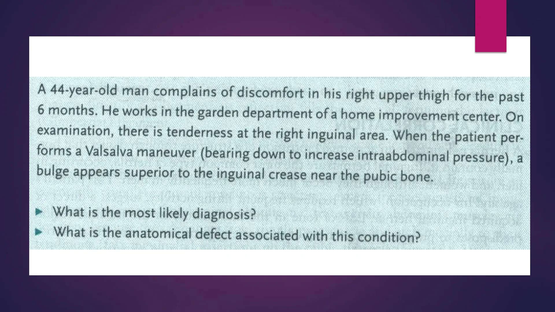
44-year-old man complains of discomfort in his right upper thigh for the past 6 months. He works in the garden department of a home improvement center. On examination, there is tenderness at the right inguinal area. When the patient per- forms a Valsalva maneuver (bearing down to increase intraabdominal pressure), a bulge appears superior to the inguinal crease near the pubic bone.
- What is the most likely diagnosis? What is the anatomical defect associated with this condition?
Descrizione del caso clinico
Uomo 44 anni Lamenta un fastidio nella parte superiore della gamba sinistra insorto negli ultimi 6 mesi Anamnesi positiva per lavoro con sforzo fisico (giardiniere)
- Indolenzimento nell'area inguinale di destra Quando il paziente esegue la manovra del torchio addominale (aumentare la pressione addominale) si nota la comparsa di una protuberanza superiormente alla piega inguinale vicino all'osso pubico7
Correlazione clinica
CLINICAL CORRELATION A hernia is defined as an abnormal protrusion of a structure through tissues that nor- mally contain it. Inguinal hernias are the most common type of hernia, occurring in men and women, although they occur much more frequently in men. This patient's age and his occupation, which requires frequent lifting activity, suggest a direct or acquired inguinal hernia. Loss of tone in the musculature in the inguinal region predisposes to progressive stretching of the parietal peritoneum into the posterior inguinal canal with repeated increased intraabdominal pressure associated with the lifting activity. If the patient were a young man or child, an indirect or congenital inguinal hernia would be a more likely diagnosis. With an indirect hernia, the pari- etal peritoneum at the deep inguinal ring exists as a fingerlike protrusion into the inguinal canal. This is the result of faulty closure of the embryonic outpouching of peritoneum into the scrotum, called the process vaginalis. Indirect inguinal hernias enter the deep inguinal ring, stretch peritoneal tissue with repeated increases in intraabdominal pressure, traverse the length of the inguinal canal, and enter the scrotum. Surgical repair of the tissue defect is indicated to prevent incarceration, infarction, and necrosis of the herniated tissue, typically a loop of small intestine.
Definizione di ernia
Un'ernia è una protrusione anormale di una struttura attraverso le fasce tissutali che fisiologicamente la contengono.
- Insorge tipicamente negli uomini adulti (ernia diretta) soggetti ad attività lavorative che spesso richiedono sforzi o sollevamento di pesi La perdita del tono muscolare della regione inguinale facilita la protrusione del viscere avvolto dal peritoneo all'interno dell'anello inguinale interno (locus di minore resistenza della parete addominale). L'ernia può insorgere anche in bambini, il tal caso viene definito ernia congenita o indiretta. E' dovuta a una deficit di chiusura del canale inguinale in cui una protrusione del peritoneo all'interno dello scroto, il processo vaginale, può favorire l'erniazione di un viscere.
Manovra del Valsalva o del torchio addominale
La manovra di Valsalva consiste in un'espirazione forzata a glottide chiusa. La manovra di Valsalva aumenta notevolmente la pressione intratoracica ed intra-addominale, favorendo tra l'altro lo svuotamento dei visceri.
Discussione
DISCUSSION The inguinal region is the junction between the lower anterior abdominal and the upper anterior thigh. It is the site at which several structures enter and exit the abdomen and therefore is an area of potential weakness in males and females. The inguinal (Poupart) ligament is an important anatomical structure and key landmark for this region. It is the thickened, rolled underedge of the inferior portion of the external abdominal oblique aponeurosis. It extends from the anterior superior iliac spine to the pubic tubercle and fuses inferiorly with the fascia lata (deep fascia) of the anterior thigh. At the pubic tubercle, the inguinal ligament continues postero- laterally on the superior pubic ramus (pectin of the pubic bone) as the pectineal (Cooper) ligament. At the point where these two ligaments are continuous and change directions, a ligamentous reflection fills the interval, forming the lacunar (Gimbernat) ligament.The lacunar ligament forms a rigid medial margin for the femoral ring, leading to the femoral canal, the site for femoral hernias (Figure 18-1).
Muscolatura e strutture anatomiche
Although the external abdominal oblique muscle and aponeurosis constitute an essentially complete musculotendinous structure (except for the superficial ingui- nal ring), the internal abdominal oblique and transversus abdominis muscles are deficient because they originate from the iliopsoas fascia and arch medially to their tendinous (falx inguinalis) insertions on the pubic tubercle (Figure 18-2). Structures enter and exit the abdomen superior to the inguinal ligament through an oblique passage known as the inguinal canal. The canal is frequently described as a tunnel, with openings, walls, floor, and so on. These boundary features are listed in Table 18-1.Two points in Table 18-1 are of anatomical and clinical significance. First, as a result of the arching of the internal oblique and transversus abdominis muscles, the posterior wall of the canal is deficient and weak, as it is formed only by the trans- versalis fascia and parietal peritoneum. However, with increased intraabdominal pressure (as in lifting, a bowel movement, etc.), these muscles contract and descend Inguinal ligament Inferior epigastric artery and vein Deep inguinal ring Rectus abdominis muscle Femoral nerve Iliopsoas muscle Hesselbach's area External iliac artery External iliac vein Femoral ring Lacunar ligament Pectineal ligament Pectineal line of os pubis Ductus deferens Abberant obturator arteryin a shutterlike fashion, thus reinforcing the posterior wall. Second, the outpouch- ing of the transversalis fascia to form the deep inguinal ring occurs immediately laterally to the inferior epigastric vessels (see Case 17 for a discussion of their course). In addition, at the medial portion of the inguinal ligament on the inte- rior of the abdominal wall, a clinically important inguinal (Hesselbach) triangle is formed by some of these structures. This triangle is formed by the inguinal ligament, inferior epigastric vessels, and lateral margin of the rectus abdominis muscle and corresponds to the area where the posterior wall of the canal is deficient because of the arching of the abdominal wall muscles described above.
Canale inguinale
In females, the inguinal canal is traversed by the round ligament of the uterus; in males, the spermatic cord (ductus deferens and associated vessels and nerves) passes through the canal. The ilioinguinal nerve is found in the canal in both sexes. The inguinal region and canal serve as the site for inguinal hernias. Although hernias occur in both sexes, they are far more common in males. There are two types of inguinal hernias: indirect and direct. Indirect or congenital inguinal hernias tend to occur in young males. During embryonic descent of the testes, an outpouchingof parietal peritoneum, the tunica vaginalis, pushes through the lower abdomi- nal wall, encountering first the transversalis fascia (thus forming the deep inguinal ring), slipping inferior to the transversus abdominis muscle, but catching the lower margin of the internal abdominal oblique muscle, and then pushing through the external abdominal oblique muscle (forming the superficial inguinal ring). The testes descend into the scrotum along the path created by the tunica vaginalis (and the gubernaculum). In normal development, this outpouching fuses and closes. If it does not fuse and close, a predisposing complete or partial path for the abnormal migration of an abdominal organ (usually small intestine) is established. The loop of small intestine would pass through the deep inguinal ring and the inguinal canal, and possibly through the superficial ring into the scrotum. By definition, indirect inguinal hernias leave the abdominal cavity lateral to the inferior epigastric vessels (through the deep inguinal ring).J
Ernie inguinali dirette
Direct inguinal hernias are also called acquired inguinal hernias because they are seen in older males and are related to strenuous activity that increases intraabdominal pressure. It is believed that with aging there is loss of tone in the abdominal musculature, and the shutterlike actions described above for the internal abdominal oblique and transverses abdominis are diminished or lost. This predisposes abdominal organs to push directly anterior through the parietal perito- neum and transversalis fascia in the inguinal triangle area and into the posterior wall of the canal. Because of the larger herniation, these hernias tend not to enter the scrotum. Direct inguinal hernias by definition leave the abdomen medial to the infe- rior epigastric vessels because these vessels form the lateral boundary of the triangle.
Passaggio dell'ernia inguinale
L'ernia inguinale diretta passa attraverso il triangolo inguinale e l'anello inguinale esterno (mediale ai vasi epigastrici inferiori) L'ernia inguinale diretta passa attraverso l'anello inguinale interno (laterale ai vasi epigastrici inferiori muscolo retto con guaina posteriore (linea semicircolare di Douglas) vasi epiga fascia trasversale inferiori `anello inguinale profondo vasi testicolari kilifaci muscolo anello femorale østert ileopsoas sinfisi del pube deferente legamento pettineo di Cooper falce inguinale e tendine congiunto legamento lacunare di Gimbernat legamento inquinale
Ernia inguinale indiretta
Ansa dell'intestino che entra nel sacco erniario Condotto deferente, vasi testicolari e ramo genitale del nervo genito-femorale all'interno del funicolo spermatico Cattura rettangolare Colletto del sacco erniario Vasi epigastrici inferiori Origine della fascia spermatica interna della fascia trasversale a livello dell'anello inguinale profondo (addominale) Peritoneo Fascia extraperitoneale Fascia trasversale Uncino che tira il muscolo trasverso dell'addome Uncino che tira il muscolo obliquo interno dell'addome Anello inguinale addominale (profondo) Fascia spermatica esterna Uncino che tira l'aponeurosi del muscolo obliquo esterno dell'addome Anello inguinale superficiale Muscolo e fascia cremasterici Fascia spermatica interna 7 Netter M.D. Sacco erniario Condotto deferente e vasi del funicolo spermatico CELSEVIER 7
Non hai trovato quello che cercavi?
Esplora altri argomenti nella Algor library o crea direttamente i tuoi materiali con l’AI.
